Friday, April 15, 2022

Justice Voice against Racism صداى عدالتخواهى عليه نژاد پرستان

 بلاخره رئيس سازمان بهداشت جهانى صداى خود را در پيوند به تبعيض عمدى #اوكراين_فرا_امتيازى بلند كرد و گفت تمركز نژادپرستان بيشتر بالاى اوكراين است تا ايتيوپيا ،

اين صدا هرچند از آدرس رياست سازمان بهداشت جهانى غير متعارف است ولى قابل قدر است؛ بويژه در جهان كه همه اى ما به عنوان يك نسل، نوع و يك نژاد در حال  نابود شدنيم، 


در هر كجاى جهان كه بروى هر كشور و هر اجتماعى و كتله اى  بزرگانى از خود دارند؛ وقتى كه اين صدا هاى عدالت طلبانه را مى شنوم دلم به كشورى مثل ما مى سوزد كه  در هر شرايطى آسيب پذير ترين، متفرق ترين و بى سرپناه و سرپرست ترينيم .


حتى همان رهبران پشتون، تاجيك ، ازبيك و هزاره كه بالاى شانه هاى هم مليتان شان سوار بودند و ازين آدرس صاحب نان و نام شدند،  صدايشان براى  دفاع از  مردم شان بر نمى آيد و اين همان مردم است كه هنوز هم بايد حتى فرزندان و نواده هاى اين رهبران را با  پمپرز هاى پُر شان بالاى شانه هاى خود بنشانند. 


وقتى كه مى بينم بنام برنامه تخليه و انتقال چه جنايات را همين بزرگان افغان انجو ها ، و رسانه ها  ، سفارت ها و نظاميان  براى مردم مستحق كشور ما اعمال كردند و خودشان اكنون از همين آدرس  تفرقه و تشنج افغانستان تا اقارب درجه بيستم خود را به كشور هاى خارجى خواستند و روز به روز امتياز خواهى بيشتر مى كنند و مردم مستحق را زير پا كردند، به مافياى افغانى در خارج كشور پى ميبرم كه با تبانى با مافيا وزارت هاى خارجه افغانستان و غربى در حال تقسيم وند و پيوند اند. 


شما ببينيد بنام اوكراين، حالا در كشور هاى اروپايى تمام مردان و پسران كشور هاى امن ديگر كه حتى براى مدتى كوتاه اوكراين رفته بودند با سوء استفاده از فرا امتيازى اوكراين به  كشور هاى اروپايى آمدند و بنام اوكراين امتياز غير قابل مقايسه اخذ مى كنند ، اينها مردان اوكراينى نيستند ، اينها از كشور هاى ديگر استند؛ اين يك پيام دارد؛ و آن اينست كه نژاد پرستى هنوز در درجه اعلى آن وجود دارد و نبايد نژاد خود را فراموش كنيم. 


در حاليكه اكنون ملت افغانستان چه در  داخل و چه در خارج فاقد ليدرشيپ سياسى است كه تا صدا بزند ، آهاى غربى ها ؛ داريد اين اشتباه قاتونى را مرتكب مى شويد، شما در بين جوامع تان جوامع كوچكتر ديگر را شكل مى دهيد، شما بايد قدر شرقى هاى غير اروپايى را بدانيد ، اكنون تمام پيشرفت بازار كار  شما از بركت مهاجرين است و چرخ شما  بالاى شانه هاى مهاجرين شرقى ( افغان، ترك، ايرانى، سورى، عراقى و افريقايى) استوار است. 


در اخير كه شما پاليسى سازان تقاعد ميكنيد ؛ نسل جوان شما كه خيلى ناچيز و راحت طلبند، كار نمى كنند، استراحت مى كنند ولى اين  نسل مهاجر شرقى است كه شما را نگاه خواهد كرد، نه راحت طلبانى ديگرى مثل اوكراين! 


جهان غرب به همان گونه كه مسول سقوط و تنزل افغانستان است ، مسول سقوط اوكراين هم است، پس اين  سبك و سنگين سازى هايتان  چيزى را به دنبال ندارد الى تشويق نژاد پرستى!.

Monday, April 4, 2022

POISE-3: Tranexamic Acid Reduces Severe Bleeding Risk in Patients Undergoing Noncardiac Surgery

 A single dose of tranexamic acid (TXA) at the beginning and end of surgery significantly reduced major bleeding in patients undergoing noncardiac surgery compared with placebo, based on findings from the POISE-3 trial presented April 2 at ACC.22 and simultaneously published in the New England Journal of Medicine.

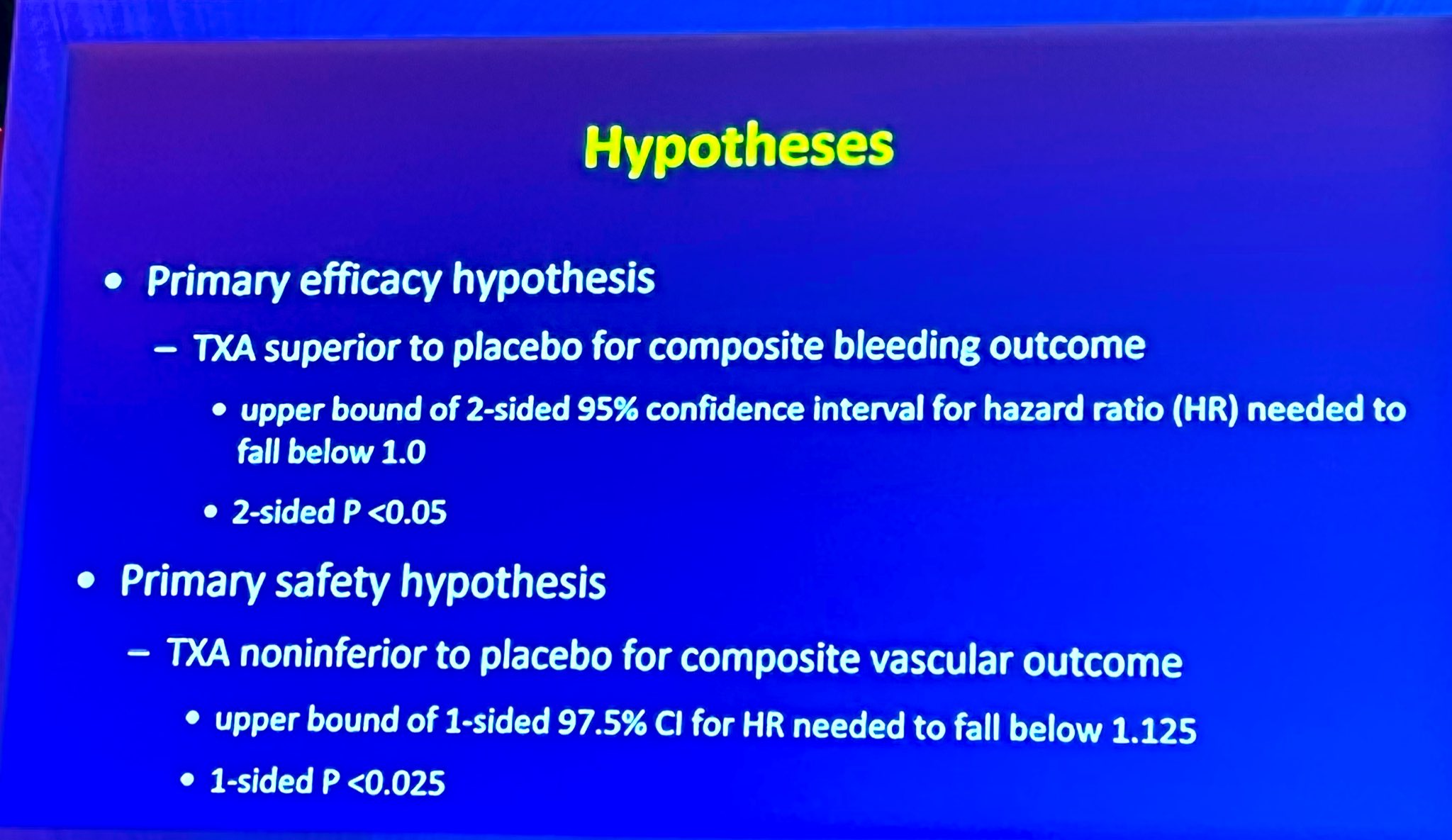
The study randomized 9,535 patients undergoing noncardiac surgery at 114 centers in 22 countries to receive TXA (1-g intravenous bolus) or placebo at the start and end of surgery. The primary efficacy outcome was a composite of life-threatening bleeding, major bleeding, or bleeding into a critical organ at 30 days. The primary safety outcome was a composite of myocardial injury after noncardiac surgery, nonhemorrhagic stroke, peripheral arterial thrombosis, or symptomatic proximal venous thromboembolism at 30 days. Patients' average age was 70 and 56% were men.


Overall results showed a composite bleeding outcome event occurred in 9.1% of those patients in the TXA group (n= 433/4,757) compared with 11.7% of patients in the placebo group (n=561/4,778 patients). Rates of adverse cardiovascular events were similar in both groups (14.2% for patients who received TXA, 13.9% for those who received a placebo). Researchers noted the benefit of TXA treatment was consistent, regardless of the type of noncardiac surgery patients received and whether patients' hemoglobin was normal or low before surgery.

"We saw an unequivocal benefit of treatment [with TXA] on preventing bleeding and blood transfusions, with no increased risk of complications," said P.J. Devereaux, MD, the study's lead author. "Given the number of people worldwide who undergo noncardiac surgery every year and the frequency of postsurgical bleeding complications, this has the potential to help a lot of patients, and I hope it will lead to a substantial change in practice."

A limitation of the study is that it did not meet its predetermined margin for determining that TXA was no less safe than the placebo. However, statistical analysis showed with a probability of more than 95% that the safety of TXA and placebo were similar. A beneficial reduction in bleeding risk must be weighed against a low probability of a small increase in the risk of a heart attack, stroke or another cardiovascular adverse event, Devereaux said.


3 April 2022

De. Nabil Paktin

Sunday, April 3, 2022

انقلاب بزرگ در سير تكامل طبابت قلبى: تداوى دارويي يا عمل جراحى براى (بيمارى ضخامت بندشى عضله قلب ): HCM

    تحقيق VALOR-HCM نشان داد كه ديگر بيماران HCM باستفاده از داروى mavacamten  ٧٧٪؜ كمتر به عمل جراحى ضرورت خواهند داشت

🚨كارديومايوپاتى هايپرتروفيك بندشى HCM يكى از علل عمده اى مرگ هاى ناگهانى قلبى در بين علل بيمارى هاى ولادى قلب نزد ورزشكاران است كه متاسفانه در كشور هاى كه ورزشكاران به گونه اى منظم مورد رد يابى بهداشتى قرار نمى گيرند ، بعد از مرگ هاى آنى ، بيمارى HCM نزد شان تشخيص مى شوند. 


🚨شيوع HCM همچنان در بين نفوس به 0.16 تا 0.29% يعنى يك نفر در بين ٦٢٥ و يا يك نفر در بين ٣٤٤ نفر واقع مى شود. ميزان مرگ و مير ساليانه آن در نزد نفوس غير ورزشكاران مصاب به ٦٪؜ مى رسد. درين بيمارى جدار هاى عضلى قلب ( خصوصا در شكل بندشى آن جدار ديوار بين القلبى ) از حد نارمل آن بسيار ضخيم تر مى شود كه در نتيجه ضربات قلب خيلى مبالغوى و قوى مى شوند ، يك نوع بندش در برابر جريان خون قلب وشريان بزرگ قلب واقع شده ،  استرخاء قلب مختل شده و مصارف انرژى قلب همچنان مختل مى شود. 


براى آن دو نوع تداوى ريشه اى تا اكنون وجود داشت كه عمل جراحى باز قلب است ؛ در آن ميخواهند از جدار هاى قلب يك قسمت عضلى قلب را قطع كنند كه تا مانع بندش پرتاب خون به دوران عمومى بدن نشود. روش دوم عمل مداخلوى نيمه جراحى است كه در لابراتوار كتيترايزيشن بالاى بيماران انجام ميشود و از طريق شرايين كرونرى يك مقدار الكل به جدار عضله قلب بيماران تزريق ميشود كه اين الكل باعث تكمش و خورد شدن همان قسمت عضله شده و مانع بندش پرتاب خون شده و اعراض بيماران در نتيجه بهبود ميابد. 


🚨اما هبچكدام اين عمل ها در نزد بيماران خالى از خطر نيست، چنانكه عمل حراحى باز قلب در يك مركز با تجربه ٦٪؜ و در مراكز بدون تجربه ١٦٪؜ خطر مرگ و مير در نزد اين بيماران را در قبال دارد 

🚨همچنان در جريان پروسيجر هاى مداخلوى نيمه جراحى، اختلاطات آن به ترتيب ؛ مرگ و مير ١.٤٪؜ ، آريتمى مداوم بطينى ٣٪؜ ،تامپوناد قلبى ٣٪؜ و ضرورت به نصب كاشت پيسميكر در نزد ٢٠٪؜ بيماران واقع مى شود. 


🚨همچنان تداوى دواى براى تخفيف اعراض وجود دارد كه هبچ كدام جزو تداوى ريشه اطلاق نمى شوند. 


🚨قبلاً يك تحقيق زير نام Explorer HCM trial در مورد شده بود كه تا ادويه جديد را منحيث تداوى ريشه اى براى كاهش حجم و يا كتله عضله قلب باعث شود كه بنابر مشكلات تخنيكى آنقدر مورد پيشانى باز جامعه طبى قرار نگرفت. ولى در آن تحقيق هم داروى Mavacamten به مقايسه گروه كه اين دارو را اخذ نكرده بودند ( پلاسيبو) موثر ثابت شد. 


🚨حالا تحقيق موثق دوم  در مورد،  زير نام VALOR-HCM صورت گرفت كه داروى Mavacamten ماواكامتين را منحيث تداوى كاهش دهنده حجم عضله قلب براى بيماران بيشنهاد كردند كه طى ١٦ هفته براى بيماران اين دارو توصيه شد. ديروز در جريان كنگره سال ٢٠٢٢ كالج كارديولوژى امريكا نتيجه اين تحقيق قناعت بخش اعلان شد. گفتنى است كه اين دوا Mavacamten باعث مى شود كه روند cross-bridge  يا بهم چسبيدن تار هاى عضلى بيشتر واقع نشده و در نتيجه قلب قويتر ضربه نزده و مرحله استرخاء وروند  مصرف انژى قلب بهبود يابد. 


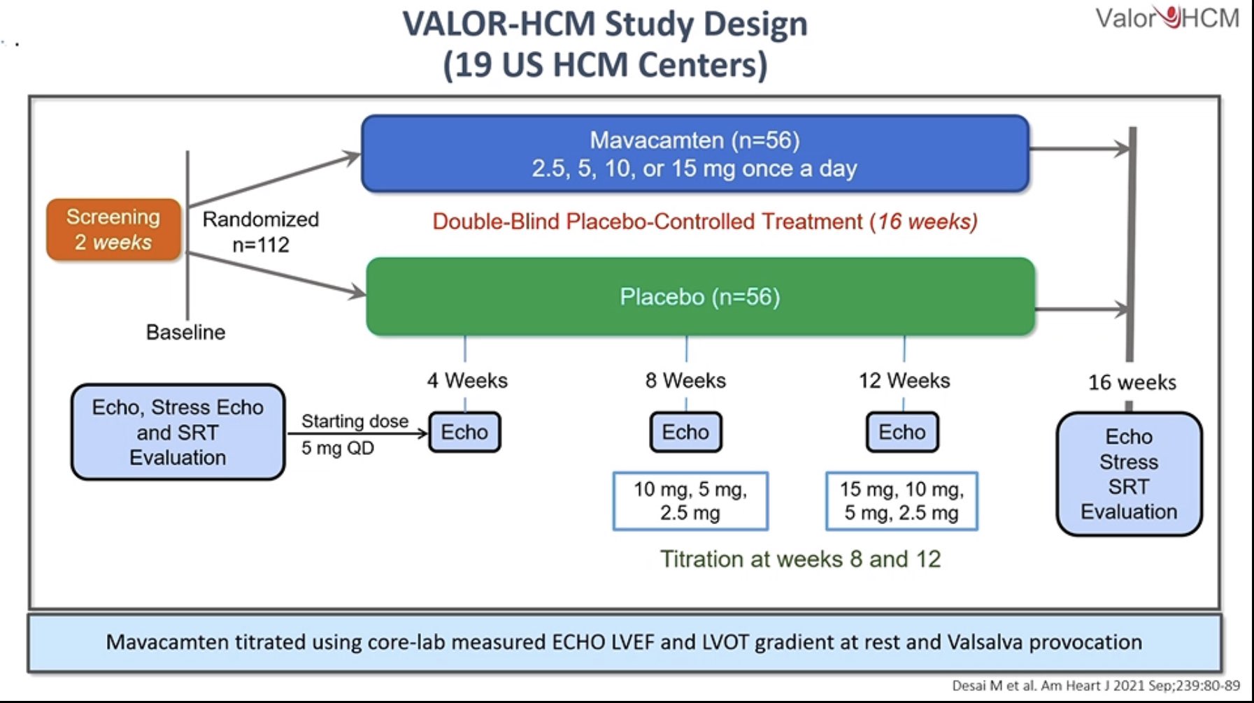
🚨درين تحقيق موثق از نوع RCT داروى mavacamten را بالاى ١١٢ بيماران  كه داراى اعراض شديد بيمارى كارديومايوپاتى بندشى هايپرتروفيك بودند در ١٩ مركز متفاوت بهداشتى مورد تجربه قرار دادند. تمام اين ببماران داراى شواهد ضخامت جدار عضله قلب به اندازه ١٥ و يا ١٣ ميلى متر و يا بيشتر از آن، تاريخچه خانوادگى HCM ، اعراض شديد علبرغم اخذ تداوى كافى دوايى بر حسب تحمل ، گراديانت قابل ملاحظه مجراى بيرونده بطين چپ (LVOT) در حال استراحت و يا تنبيه حدود ٥٠ يا بيشترِ به ميلى متر ستون سيماب و يك EF بيشتر از ٦٠٪؜  داشتند. 


🚨محققين به طور روند تصادفى بيماران را در دو گروه كه دارو را اخذ مى كنند و بيماران كه اين دارو را اخذ نميكنند نقسيم كردند و براى مدت ١٦ هفته مورد رد يابى قرار دادند. اولين عواقب تخمينى تحقيق  بعد از ١٦ هفته تداوى ضرورت به عمل جراحى و يا مداخلوى قلمداد شده و عواقب تخمينى دوم تحقيق عبارت از كاهش گراديانت از طريق مجراى ببرونده بطين چپ ،كاهش در  يك و يا بيشتر از كلاس هاى NYHA , بهبودى كيفيت زندگىً، كاهش سويه NT ProBNp و كاهش در سويه تروپونين I. بودند.


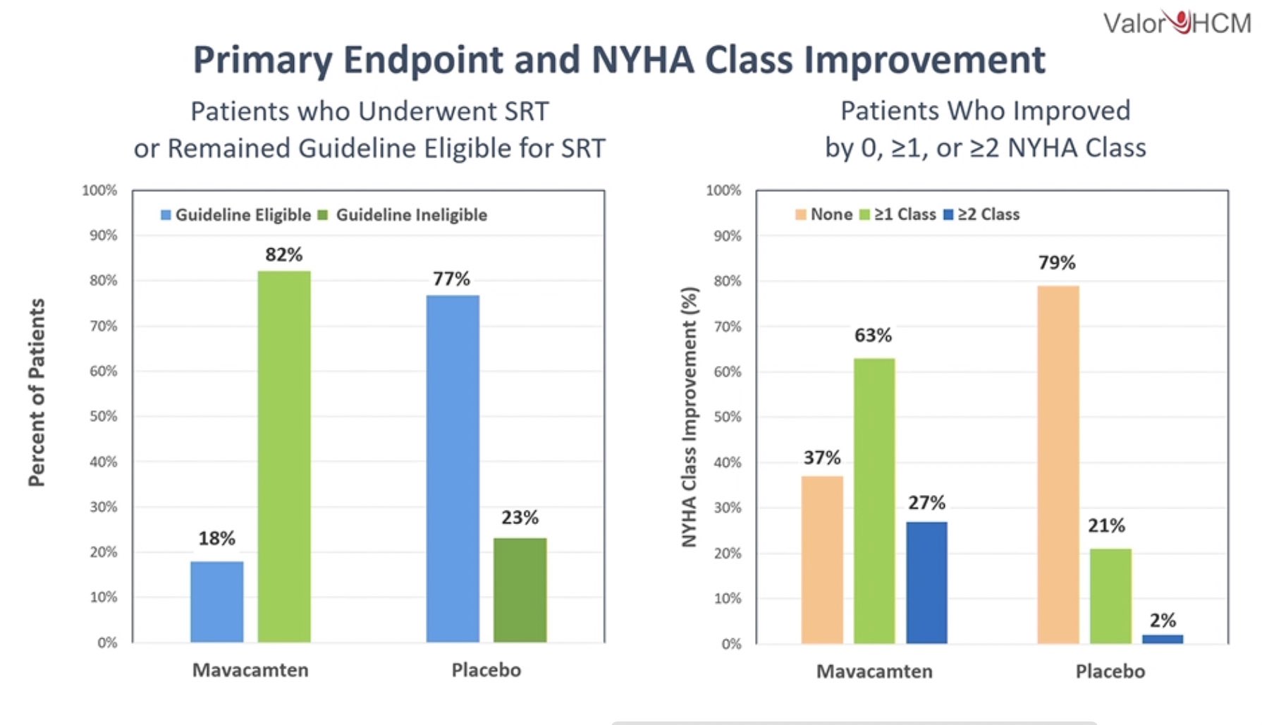
🚨در نتيجه رد يابى ١٦ هفته اى ١٨٪؜ بيماران به جراحى و مداخلات اصلاحى ضرورت پيدا كردند و ٧٧٪؜ بيماران به جراحى ضرورت نيافتند و در تمام شماره هاى عواقب تخمينى دومى تحقيق بهبودى مطلوب واقع شده كه هيچ نوع اختلاطى در مورد گزارش نشد. 


🚨اين اولين روش تداوى داروي است كه ضرورت بيماران به عمل جراحى قلبى را كاهش مى دهد.

به تاسى ازين تحقيق اكنون اداره دوا و غذاى آمريكا الى ختم ماه اپريل اجازه رسمى خود را براى استعمال اين دارو در نزد اين گروه بيماران اعلان خواهد كرد. 

🚨درين مورد تحقيقات بيشتر براى مدت زمان بيشتر ضرورت است كه تا مصونيت بيشتر اين دارو را بالاى بيماران در استعمال طولانى مدت اين دارو ، مورد ارزيابى قرار  دهد. 

گفتنى است كه يكى از عوارض جانبى موقتى اين دارو پايين افتادن EF بيماران بود كه با قطع موقت و ادامه دوباره آن اين مشكل حل شد.


يادداشت: بعدا ً در كتب چاپ آينده ازين تحقيق. تذكار بعمل خواهد آمد و درج تداوى هاى داروى كتب  خواهند شد، از همين رو خواندن تخقيقات ما را جلو تر و به روز تر از كتب مى سازند.


تبصره و نوشته بر تحقيق: #دكتور_نبيل_پاكطين

٣ اپريل ٢٠٢٢

اعلان تحقيق٢ اپريل. ٢٠٢٢

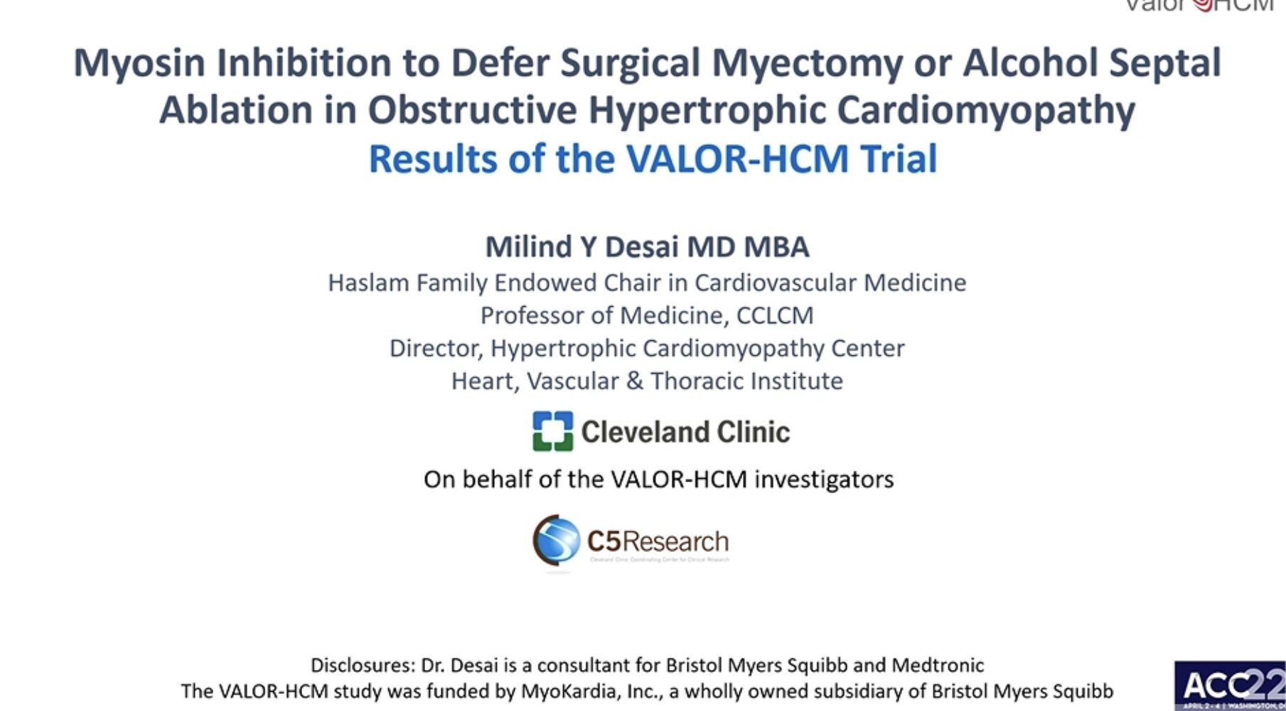
VALOR-HCM: Mavacamten Significantly Reduces Need For Surgical Intervention in Patients With Obstructive HCM


 Use of mavacamten to treat patients with obstructive hypertrophic cardiomyopathy (HCM) who were referred as candidates for invasive septal reduction therapy (SRT), significantly reduced the need for the surgical intervention at 16 weeks, according to results from the VALOR-HCM trial presented April 2 during ACC.22.

The randomized, double-blind, placebo-controlled study evaluated the use of mavacamten in a total of 112 adults with symptomatic obstructive HCM who were referred to an HCM expert at 19 HCM centers. All patients had documented HCM with maximum septal wall thickness ≥15 mm or ≥13 mm; family history of HCM; severe symptoms despite maximally tolerated medical therapy; dynamic left ventricular outflow tract (LVOT) gradient at rest or with provocation ≥50 mmHg; and documented left ventricular ejection fraction ≥60%.


Researchers randomly assigned each patient to receive mavacamten or a placebo for 16 weeks. The primary endpoint was a composite of patient decision to proceed with SRT or continue to meet 2011 ACC/AHA guideline eligibility for SRT after 16 weeks.


At 16 weeks, only 18% of patients on mavacamten were still eligible for SRT, compared with 77% of those on placebo. Researchers also observed significant improvements across all secondary endpoints, including reduction in resting LVOT gradient; ≥1 class improvement in NYHA Class; improvement in KCCQ-clinical summary score; reduction in N-terminal pro-B-type natriuretic peptide; and reduction in troponin I. No patients experienced serious adverse cardiac events.

"These were very symptomatic, sick patients who were on maximally tolerated medical therapy and were faced with the decision of whether or not to have septal reduction therapy to relieve the obstruction. In taking this drug, they got a whole lot better across the board," said Milind Y. Desai, MD, MBA, FACC, the study's lead author. "This is really the first pharmacotherapy that offers a viable medical option for people with obstructive HCM short of needing a procedure."

Desai and colleagues note that additional data is needed to assess the durability of improvement in SRT eligibility over longer time periods. "We are hoping this study will also draw much-needed attention to HCM," said Desai. "This disease is woefully underrecognized. There are not enough HCM experts and not enough proven medical therapies, so there are many opportunities to do better."

Mavacamten is currently under review with the U.S. Food and Drug Administration for use in obstructive HCM, with a decision expected at the end of April.


ACC 2022


Prepared by: Dr. Nabil Paktin,


Saturday, March 12, 2022

Review of anticoagulation in patients with Covid19 Key points:

 The following are key points to remember from this review on anticoagulation in patients with coronavirus disease 2019 (COVID-19):

  1. COVID-19 has led to unprecedented morbidity and mortality with >200 million cases and 4 million deaths worldwide. Apart from acute respiratory distress syndrome, COVID-19 is associated with thromboembolic disease.
  2. Viral entry through the endothelium may cause inflammation and vascular injury. Mononuclear cell activation can also trigger cytokine release and cytokine storm. Collectively, these lead to a prothrombotic state for patients with COVID-19. Other mechanisms may also contribute, including complement activation and anti-phospholipid antibodies.
  3. COVID-19 coagulopathy is categorized by laboratory abnormalities including increased levels of fibrinogen and D-dimer, mild prolongation of prothrombin time or activated partial thromboplastin time, and mild thrombocytopenia. Patients admitted with COVID-19 who have elevated D-dimer or troponin levels are associated with worse outcomes, including mortality.
  4. The incidence of venous thromboembolism in patients hospitalized with COVID-19 was 17% in a meta-analysis of 49 studies. The rate was higher in patients with critical illness (27.9%) as compared to those who were not critically ill (7.1%).
  5. Clinicians and researchers have explored the role of antithrombotic therapy to prevent COVID-19–associated thromboembolism. This includes use of standard prophylactic-dose anticoagulation, intermediate-dose anticoagulation, and therapeutic-dose anticoagulation given for prophylactic purposes.
  6. Several large observational studies have suggested that prophylactic-dose enoxaparin was associated with lower rates of intubation and death. However, the role of higher-dose prophylactic anticoagulation in preventing poor outcomes is not clear from observational studies.
  7. Two large trials have explored the use of anticoagulation outside of the hospital setting for patients with COVID-19. The MICHELLE trial compared rivaroxaban 10 mg daily for 35 days after hospital discharge with placebo and found a 67% relative risk reduction in their primary outcome without an increased risk of bleeding. Conversely, the ACTIV-4B study compared apixaban versus placebo in ambulatory patients with mild COVID-19 but was ended early due to a very low rate of thromboembolic events that did not meaningfully differ between treatment arms.
  8. Several trials have compared intermediate- or treatment-dose anticoagulation with standard prophylactic-dose anticoagulation in hospitalized patients with COVID-19. The multiplatform trial included 2,219 patients hospitalized without critical illness and found an increase in the number of organ support-free days with treatment-dose anticoagulation. In a concurrent trial of 1,098 critically ill patients in the multi-platform trial, treatment-dose anticoagulation did not increase the number of organ support-free days as compared to standard prophylaxis.
  9. In the HEP-COVID trial of 257 patients hospitalized with COVID-19 who required oxygen support and other risk factors, therapeutic-dose enoxaparin led to lower rates of thromboembolism and death as compared to standard-dose thromboprophylaxis. Similarly, in the RAPID trial of 465 patients with moderately ill COVID-19 and elevated D-dimer, therapeutic-dose heparin led to numerically (but not statistically) lower rates of death, intensive care unit (ICU) admission, and mechanical ventilation as compared to standard prophylactic-dose heparin.
  10. The authors are conducting a large-scale, prospective, multicenter, open-label randomized clinical trial of patients hospitalized with COVID-19 who will be randomized to receive prophylactic-dose enoxaparin, therapeutic-dose enoxaparin, or therapeutic-dose apixaban. The primary effectiveness outcome will be a composite of all-cause mortality, requirement for intensive care, systemic thromboembolism, or ischemic stroke within 30 days.

Geoffrey D Barnes MD., FACC

Review of renal compression in heart failure

 The following are key points to remember from this state-of-the-art review on renal compression in heart failure (HF):

Key points

  1. Previous studies have linked poor renal function in HF to both decreases in perfusion (due to low cardiac output) and increases in renal congestion (due to increases in central venous pressure). The latter is believed to play a significant role in worsening renal function (WRF) in HF.
  2. The authors hypothesize that three important mechanisms, alone or in combination, may contribute to renal congestion in HF: increased intracapsular, perirenal, and intra-abdominal pressures. These increases in pressure can lead to compression of the kidney and renal vasculature.
  3. Increased intracapsular pressure: As central venous pressures in HF increase, this leads to an increase in interstitial fluid in the renal parenchyma. Given that the kidneys are surrounded by a rigid capsule, volume expansion does not occur, which means increased interstitial fluid causes an increase in intracapsular pressure. This causes compression of renal veins, glomeruli, and tubules, which subsequently may lead to WRF.
  4. Increased perirenal pressure: Just outside of the renal capsule but within the renal fascia is the perirenal space, comprised mostly of adipose tissue. While not fully understood, increases in perirenal adipose tissue, as well as renal sinus fat within the kidney, may lead to renal vein compression and exacerbation of WRF in an already congested kidney.
  5. Increased intra-abdominal pressure: In patients with significant HF, increases in intra-abdominal pressure can be a result of many processes, which include development of ascites or retention of fluid in the splanchnic system. This leads to compression of renal vasculature and can contribute to WRF in HF. A similar effect can be seen in patients with significant obesity leading to increased intra-abdominal pressure.
  6. The authors suggest that renal decapsulation in HF patients may possibly lead to improved renal outcomes. Data from rat and piglet animal models suggest that decapsulation reduced renal tubular damage and ischemic acute kidney injury.
  7. The authors introduce the “renal tamponade” hypothesis to explain why significant WRF is present when central venous pressures are high in HF. Intra- and extra-renal compression of the kidney and renal vasculature through the above three mechanisms are major contributors.
  8. More research is needed to better define these mechanisms and to explore the possible benefit of renal decapsulation in managing WRF in HF.


Marty Tam MD., FACC,

6 March 2022


Review on LV Thrombus Following MI

 The following are key points to remember from this state-of-the-art review on left ventricular (LV) thrombus following myocardial infarction (MI):

  1. LV thrombus after acute MI (AMI) has declined significantly since the introduction of reperfusion therapy. The current estimate is that LV thrombus occurs in up to 6.3% of patients with ST-segment elevation MI (STEMI) and 19.2% of patients with anterior wall STEMI complicated by LV ejection fraction <50%.
  2. The imaging modality to detect LV thrombus greatly impacts the frequency of detection. Transthoracic echocardiogram (TTE) is inferior to cardiac magnetic resonance (CMR) imaging for detecting small, laminar LV mural thrombus. In fact, the sensitivity of TTE is only 29%, while the specificity is 98% as compared to CMR.
  3. CMR is the optimal imaging modality for diagnosis of LV thrombus. It maintains a sensitivity of 82-88% and specificity approaching 100% compared to surgical and/or pathological confirmation. Use of late gadolinium enhancement improves thrombus detection over cine CMR.
  4. Thromboembolic events caused by LV thrombi can be devastating. The risk of thromboembolism is most closely related to thrombus mobility and protrusion as described on imaging. The rate of thromboembolism varied between 3% in patients with consistently therapeutic anticoagulation and 19% in patients with poorly controlled anticoagulation.
  5. Most LV thrombi can be detected by imaging within 2 weeks of AMI. High-risk patients without LV thrombus on early imaging (e.g., within 48 hours after AMI) should be reimaged 2 weeks after the acute event.
  6. Virchow’s triad, which outlines the pathophysiology of thrombosis formation, applies to LV thrombus following AMI. Blood stasis from LV dysfunction and apical/anterior aneurysms is complicated by a hypercoagulable state from inflammation, elevated fibrinogen and neutrophils, platelet aggregation, and clotting cascade activation. This is further potentiated by tissue injury impacting the subendothelial tissue and exposing collagen.
  7. Therapies to prevent and treat LV thrombus following AMI also target Virchow’s triad. Guideline-directed medical therapy addresses issues of blood stasis, while anticoagulation addresses hypercoagulability and reperfusion therapy ± anti-inflammatory therapy addresses tissue injury.
  8. Prophylactic anticoagulation may be considered for patients with STEMI and anterior apical akinesis or dyskinesis. The 2013 American College of Cardiology/American Heart Association STEMI guidelines recommend use of a vitamin K antagonist (VKA) with a lower international normalized ratio (INR) target of 2.0-2.5. However, no prospective trial has examined the role of anticoagulation plus antiplatelet therapy in the modern percutaneous coronary intervention era.
  9. For patients who have been diagnosed with LV thrombus following AMI, studies have produced conflicting results regarding the safety and efficacy of VKA versus direct oral anticoagulants (DOACs). The authors recommend the use of VKA with goal INR 2-3. DOAC should be used if VKA cannot be tolerated.
  10. The authors recommend that repeat imaging be obtained after 3 months of therapy. If the LV thrombus has resolved, anticoagulation can be discontinued and dual antiplatelet therapy continued per management of AMI. If the LV thrombus is persistent, anticoagulation should continue with repeat imaging every 3 months. Once anticoagulation has been discontinued, repeat imaging 3 months later is advised.


Geoffrey D. Barnes, MD, MSc, FACC

10 March 2022