
Sunday, December 18, 2016
Monday, December 5, 2016
How a venous/coronary artery bypass graft fails ? (What is neointimal hyperplasia ) ?
The main limitation of coronary artery bypass
grafting (CABG), when the saphenous vein is used as
a conduit, is poor long-term vein graft patency. Fiveyear
failure rates are 30–50% and have remained unchanged
despite rapid development of pharmacological
treatments and technologies.
Perhaps the most important change following vein grafting is the exposure of saphenous vein to the arterial circulation. In the venous circulation saphenous vein is subjected to low pressure, non-pulsatile flow and a shear stress of around 0.2 dyne/cm2
Following grafting the vein is exposed to high pressure, pulsatile flow and a shear stress of approximately 3-6 dynes/cm 2. In addition to increased shear stress the vein is subjected to a variety of other new haemodynamic forces, including radial and circumferential deformation.

The another main reason for vein graft occlusion, especially in the mid-term, is neointimal hyperplasia (NIH).
Following vein grafting there is rapid deposition of leukocytes, platelets and other blood components. . These accumulating cells and blood components may have an important influence on the later development of intimal hyperptasia.In the experimental situation inhibiting leukocyte accumulation using an antibody to CD4 results in a reduction in the development of intimal hyperplasia,
while reduction in platelet aggregation using an antibody to GP IIb-IIIa has been shown to reduce the incidence of restenosis following coronary angioplasty in vivo.
Monocytes is also playing important role. Studies of excised vein graft stenoses has demonstrated an abundance of proliferating monocytes and macrophages in the intima of these lesions.
Leukocytes can release cytokines, oxygen-derived free radicals and lysosomal proteinases, which, by direct effects on smooth muscle cells and also modulation of endothelial products, e.g. inactivation of nitric oxide, may influence smooth muscle cell proliferation and migration.
Similarly deposited platelets release smooth muscle mitogens, such as platelet derived growth factor, which encourage smooth muscle cell proliferation and migration into the intima.
Studies in cultured cells have demonstrated that haemodynamic forces have an important influence on the endothelial expression of molecules controlling leukocyte and platelet adhesion. Thus it is tempting to explain the association between haemodynamic forces and vein graft thickening by their effect on the accumulation of blood elements.
It is well-known that aortocoronary grafts fashioned from internal mammary artery or radial artery are much more durable than saphenous vein grafts and it is of note that SMCs derived from internal mammary artery proliferate less than SMCs from saphenous vein.
There are significantly higher activity of phosphatase and tensin homolog (PTEN) in the smooth muscle cells of the internal mammary artery than in the saphenous vein.
In summary one can say that :
In vein-graft failure various factors pathophysiologiclly are involved , including PTEN, matrix metalloproteinases, and tissue inhibitor of metalloproteinases, in uncontrolled proliferation and migration of smooth muscle cells towards the lumen, and invasion of the graft conduit.

Sunday, November 27, 2016
Can antiplatelet agent be considered as an alternative to OAC in SPAF? Can we combine the Oral anticoagulants with anti-platelets in SPAF?
Can antiplatelet agent be considered as an alternative to
OAC in SPAF?
In terms of stroke prevention in AF, the bottom line is
effective stroke prevention means oral anticoagulation therapy and these days
it can either mean a NOAC (non vit. K oral anticoagulant) or Vit.K antagonist (VKA)
e.g. Warfarin because that is where the evidence is clearly there which shows
that OAC harpy prevents stroke. Aspirin or anti-platelet therapy had been
tested in SPAF (stroke prevention in Atrial fibrillation), and the evidence
suggests no significant benefits, there is however evidence of harm i.e.
increase in the risk of both major and intracranial bleeding. NICE guidelines
in UK, in 2014 which also undertakes a cost effectiveness analysis stated that
not only is aspirin ineffective but it is actually not safe and certainly not cost
effective. In net clinical benefits for aspirin in SPAF is essentially neutral
or trending towards harm. In short, aspirin mono-therapy should be used as Mono-therapy
in SPAF.
Can we combine the Oral anticoagulants with anti-platelets
in SPAF?
Anti-platelet therapy can be combined with oral anticoagulant
therapy essentially in a situation of the patient with AF possesses ACS or
undergoes coronary intervention including coronary stenting. In patient with
stable vascular disease essentially in majority of patients with AF there is no
demonstrated benefit to add anti-platelet therapy to oral anticoagulant therapy
because the available data shows that there is no benefit in terms of stroke reduction,
morality or myocardial infarction, however, what you do see is a significant
increase in major bleeding as well as significant increase in intracranial
bleeding when anti platelet therapy is combined with oral anticoagulation.
So in short do not combine anti-platelet therapy and oral anticoagulant
therapy in majority of patients of AF as there is little evidence of benefit,
there is certainly strong evidence of harm in these patients.
This combination therapy should be reserved when there is a
necessity to have associated anti-platelet therapy most commonly after an ACS
or a coronary stent intervention.
Tuesday, October 25, 2016

Saturday, October 22, 2016

Friday, September 30, 2016
What is intra cardiac blood cyst ?
Blood cyst in the heart is a very rare finding and was first reported by Elasser in 1844. The cysts are most commonly present on the supporting structures; atrioventricular valves, accounting for 96% of the cysts in infants, and are less often present on pulmonary and aortic valves.
Histologically,
it is thin-walled and normally lined by cobblestone-shaped endothelial cells
and does not contain any tumorous cells.
Blood
cysts are often asymptomatic, small and congenital. The cysts regress
spontaneously in most patients and are consequently rare in adults, there are
some cases reported in contrast. Cyst
growth potential complications include valve dysfunction, left ventricular
outflow tract obstruction, and embolic stroke have been documented.
In differential
diagnosis primary cystic tumor such as
hemangioma or myxoma should be taken into account and the right-sided cystic
mass includes
aneurysmatic
atrioventricular septum, cavitating thrombus, abscess formation as a process of endocarditis, hydatid cyst, and blood cyst.
However,
absence of intracystic calcification, homogenous pattern of cystic
fluid, relation
to the tricuspid valve, and clinical history strongly suggested a blood cyst in
our patient.
Echocardiography indicated the cystic nature of the tumor which is highly mistaken with cardiac hydatidosis. However, cardiac MRI was important for its diagnosis.
Echocardiography indicated the cystic nature of the tumor which is highly mistaken with cardiac hydatidosis. However, cardiac MRI was important for its diagnosis.

Hydatid cysts
exhibit a different behavior under MRI, being a
a round
homogeneous image is observed with signs of bleeding (iso- or hyperintense in
T1 and iso- or hypointense in T2) with no uptake of IV contrast media, which
indicates its hematic and cystic nature
Because of the
cyst’s location, a myxoma could be suspected, but myxomas tend to be
heterogeneous, and although some may exhibit a more homogeneous behavior, they
always exhibit contrast uptake, being solid lesions.
A chronic
thrombus may have similar intensity in T1 and T2, but its round morphology, its
well-defined margins, the presence of a tiny pedicle, and its cystic nature as
revealed by MRI and echocardiography do not support this diagnosis.
Although a
cardiac blood cyst is a very rare finding, it can
be diagnosed
using cardiac MRI and it should be included in
the
differential table of masses inside heart cavities.
There are several purposed mechanisms for formation of cystic mass ,however, it is believed that invagination at crevices of the valve surface into stroma by high ventricular pressure may result in blood-filled cyst formation. Subsequently, the mouths of the crevices may fuse to form a closed cyst.
The
followings are hypotheses :
The first is that blood
cysts are formed during valve development as a result of blood being pressed
and trapped in crevices that are later sealed off.
The second hypothesis is
that blood cysts are the result of hematoma formation in the subvalvular region
secondary to the occlusion of small vascular branches of end arteries due to
inflammation, vagal stimulation, anoxia, or hemorrhagic events.
The third hypothesis
involves possible heteroplastic changes in the tissue that comes from primitive
pericardial mesothelium.
The fourth and fifth
hypotheses are that these blood cysts simply represent ectatic or dilated blood
vessels in the valve or that they represent angiomas.
However, there is still
no consensus regarding the development of blood cysts.
Dencker et al suggested
that a conservative approach in asymptomatic patient with minor cyst, and
surgical resection should be considered if symptoms exist or if the cysts lead
to any cardiac dysfunction.
References
1)
Michelena HI, Mulvagh SL, Schaff HV, Enriquez-Sarano ML, Klarich
KW. A heart-shaped mass inside a heart: echocardiographic diagnosis, pathology, and surgical repair of a
flail tricuspid valve caused by a large blood-filled cyst. J Am Soc Echocardiogr 2007;20:771.e3–6.
2)
Jose VJ, Gupta SN, Jose S, Chacko B, Abraham PK, Abraham OC et al. Blood-filled cysts of heart. Indian Heart J 2004;56:174–5.
3)
Shing M, Rubenson DS.
Embolic stroke and cardiac papillary fibroelastoma. Clin Cardiol 2001;
24:346-7.
4)
Prasad A, Callahan MJ, Malouf JF. Acquired right atrial blood
cyst: a hitherto unrecognized complication of cardiac operation. J Am Soc Echocardiogr
2003; 16: 377–378
5)
López-Pardo F, López-Haldón J, Granado-Sánchez C, Rodríguez- Puras
MJ, Martínez-Martínez A. A heart inside the heart: blood cyst of mitral valve.
Echocardiography 2008;25:928-30.
6)
7)
Kuvin J, Saha P, Rastegar H, Salomon RN, Pandian N, Denofrio D. Blood
cyst of the mitral valve apparatus in a woman with a history of orthotopic
8)
Dencker M, Jexmark T, Hansen F, Tydén P, Roijer A, Lührs C.
Bileaflet blood cysts on the mitral valve in an adult. J Am Soc Echocardiogr 2009;22:1085.e5-8.
Sunday, August 14, 2016
A complex case of PCI ( Rota-stenting strategy for heavily calcified coronary lesions ) ,
A journal published article , written by Dr.Nabil Paktin


Saturday, July 9, 2016
What is Electrical alternans vs. Pseudo-electrical alternans and pseudo literature reports ?
Electrical alternans is a broad term that describes alternate-beat variation in the direction, amplitude, and duration of any component of the ECG waveform (ie, P, PR, QRS, R-R, ST, T, U)
It was first recognized by Hearing in 1909 and further characterized by Sir Thomas Lewis in 1910 as occurring “either when the heart muscle is normal but the heart rate is very fast or when there is serious heart disease and the rate is normal.”
Kalter and Schwartz first identified electrical alternans on surface ECG in 1948
Electrical alternans must be distinguished from mechanical alternans (eg, pulsus alternans), although both may coexist
The pathophysiologic mechanisms that cause electrical alternans can be divided into 3 categories:
-Repolarization alternans (ST, T, U alternans)
-Conduction and refractoriness alternans (P, PR, QRS alternans)
-Alternans due to cardiac motion

Electrical Alternans Associated with cardiac motion is due to alternation in the position of the heart with relation to recording electrodes.
The most common underlying disorder is an enlarged pericardial sac; however, not all pericardial effusions cause electrical alternans.
The presence of pericardial disease and total electrical alternans (P, QRS, and T wave) frequently suggests cardiac tamponade, but total electrical alternans is seen in only 5-10% of patients with cardiac tamponade.
Heart movement in patients with hypertrophic cardiomyopathy also may result in electrical alternans of this type .
Whenever what appears to be electrical alternans is not due to a large pericardial effusion, then pseudoelectrical alternans should be considered. Pseudoelectrical alternans is due to alternation in axis or amplitude because of events that alter conduction and do not alter the physical orientation of the heart.
In 1978, Klein, Segni and Kaplinsky coined the term ‘pseudoelec- trical alternans’ in a case report of intermittent left anterior hemiblock, in which the axis shifted every other beat due to the development of alternating normal and then leftward axis shift, presumably related to procaina- mide therapy.
Unfortunately, some literature defining interchangeably as true electrical alternans is a repolarization or conduction abnormality of the Purkinje fibers or myocardium.
Electrical alternans due to cardiac motion is effectively artifact, as the heart swings in relation to the chest wall and electrodes, with a period twice that of the heart rate. However, tamponade related electrical alternans is the true one .
Electrical alternans due to cardiac motion is effectively artifact, as the heart swings in relation to the chest wall and electrodes, with a period twice that of the heart rate. However, tamponade related electrical alternans is the true one .
Exclusively in Dr.Nabil Paktin Cardiology Notes
Friday, July 8, 2016
Time course and hemodynamics of Mitral stenosis ( MS) causing Pulmonary arterial Hypertension ( PAH)
Mitral stenosis occurs
Left atrial pressure rise
Left atrium enlarges
Cephalization
PIE
PAH develops
PVR increases
RV enlarges
Pulmonic regurgitation develops
Tricuspid annulus dilates
Tricuspid insufficiency
RV failure

 Please Click over image for a better resolution
Left atrial pressure rise
Left atrium enlarges
Cephalization
PIE
PAH develops
PVR increases
RV enlarges
Pulmonic regurgitation develops
Tricuspid annulus dilates
Tricuspid insufficiency
RV failure


Saturday, July 2, 2016


Sunday, June 19, 2016
Echogenic Foci ( bright spot in the baby's heart) , Is it normal or abnormal ?


References
1. Petrikovsky BM, Challenger M, Wyse LJ. Natural history of echogenic foci within ventricles of the fetal heart. Ultrasound Obstet Gynecol. 1995;5:92-94.
2. Brown DL, Roberts DJ, Miller WA. Left ventricular echogenic focus in the fetal heart: pathologic correlation. J Ultrasound Med. 1994;13:613-616.
3. Tennstedt C, Chaoui R, Vogel M, Goldner B, Dietel M. Pathologic correlation of sonographic echogenic foci in the fetal heart. Prenat Diagn. 2000;20:287-292.
4. Dildy GA, Judd VE, Clark SL. Prospective evaluation of the antenatal incidence and postnatal significance of the fetal echogenic cardiac focus: a case-control study. Am J Obstet Gynecol. 1996;175:1008-1012.
5. Barsoom MJ, Feldman DM, Borgida AF, Esters D, Diana D, Egan JF. Is an isolated fetal cardiac echogenic focus an indication for fetal echocardiography? J Ultrasound Med. 2001;20:1043-1046.
6. Lamont RF, Havutcu E, Salgia S, Adinkra P, Nicholl R. The association between isolated fetal echogenic cardiac foci on second-trimester ultrasound scan and trisomy 21 in low-risk unselected women. Ultrasound Obstet Gynecol. 2004;23:346-351.
7. Simpson JM, Cook A, Sharland G. The significance of echogenic foci in the fetal heart: a prospective study of 228 cases. Ultrasound Obstet Gynecol. 1996;8:225-228.
8. Bradley KE, Santulli TS, Gregory KD, Herbert W, Carlson DE, Platt LD. An isolated intracardiac echogenic focus as a marker for aneuploidy. Am J Obstet Gynecol. 2005;192:2021-2026; discussion 2026-2028.
Thursday, June 9, 2016
Diabetes is not CAD equivalent anymore
Diabetes and Prior Coronary Heart Disease are Not Necessarily Risk Equivalent for Future Coronary Heart Disease Events
The prevalence and burden of diabetes mellitus remains high.After Haffner et al.reported that adults with diabetes had the same risk for future myocardial infarction (MI) as adults with previous MI and without diabetes, the Adult Treatment Panel (ATP) III guidelines in 2001 recommended that all individuals with diabetes be considered as “Coronary heart disease (CHD) risk equivalent”.However, the latest 2013 ACC/AHA assessment of risk guidelines considers diabetes as only one of the many variables in its risk assessment equation.
The assertion that all patients with diabetes are CHD equivalent has been controversial.Existing evidence is based on relatively small studies with various limitations.
Some studies were limited to a single gender, while others were based on self-reported diagnosis of diabetes.
Some lacked the ability to adjust for important confounding risk factors.Most of the studies have comprised cohorts from the 1990s, and only a few studies have been able to evaluate the impact of the duration of diabetes. There is also a paucity of data among relatively young (30–40 years) patients with diabetes. For all these reasons, updated evidence from a contemporary population is needed to inform our understanding of CHD risk in diabetes patients

Based on a recent examination of the 2013 ACC/AHA Pooled Cohort Equation, the inclusion of diabetes in the scoring criteria rather than considering diabetes as an automatic CHD equivalent led to important differences in predicted risk that might influence decision-making in younger patients with diabetes.
The recent study expressed as follow:
-Individuals with diabetes alone had significantly lower risk of CHD across all age and sex strata compared to those with CHD alone (12.2 versus 22.5 per 1000 person-years).
-The risk of future CHD for patients with a history of either DM or CHD was similar only among those with diabetes of long duration (≥10 years) to can call diabetes equivalent.
-Not all individuals with diabetes should be unconditionally assumed to be a risk equivalent of those with prior CHD.

Abdominal migraine in adults , میگرن بطنی نزد بزرگسالان
هزاران بیمار روزانه از اثر درد های شکم به مراکز صحی مراجعه می کنند، بعضی ها بدون تشخیص به خارج کشور میروند و حتی در خارج مورد بهره برداری نقدی قرار می گیرند بنام های چک عمومی بی نتیجه بر میگردند ولی درد شکم همان درد است ، و برخی دیگر با تصادفات تشخیصیه مورد عملیات جراحی قرار می گیرند در حالیکه به هیچ تشخیص نهایی نرسیده اند .
میگرن یا نیم سری که همانا درد های حملوی سردردی است دیگر نمیتواند تنها در جغرافیای سر منحصر بماند ، میگرن بطنی در ابتدا منحیث مشکلات کودکان گزارش شده بود ولی به گونه شاید آن هرگز داخل تطبیقات روزانه و شنیده نشد . علیرغم لوایح تشخیصیه متنوع ، تشخیص میگرن بطنی فوق العاده مشکل است چون اکثراً توسط معاینات متممه تشخیص نمیشوند .
میگرن یا نیم سری که همانا درد های حملوی سردردی است دیگر نمیتواند تنها در جغرافیای سر منحصر بماند ، میگرن بطنی در ابتدا منحیث مشکلات کودکان گزارش شده بود ولی به گونه شاید آن هرگز داخل تطبیقات روزانه و شنیده نشد . علیرغم لوایح تشخیصیه متنوع ، تشخیص میگرن بطنی فوق العاده مشکل است چون اکثراً توسط معاینات متممه تشخیص نمیشوند .
زمانیکه تمام معاینات هضمی و بطنی نارمل آید ، منحیث تشخیص افتراقی و نهایی ، میگرن بطنی باید فراموش نشود .
یکی از نکته های که ما را میتواند به طرف تشخیص میگرن بطنی هدایت کند هما ربط درد شکم با سر دردی است ولی برعکس آن هم میتواند واقع شود که همانا درد های مکرر شکم باعث سردردی های میگرنی در چند سال آینده میشود . تداوی میگرن بطنی مشکل است چون اکثراً بیماران در برابر تداوی متعارف چون مسکن ، انتی اسید و ضد تهوع مقاومت نشان می دهند .
تداوی دوایی ضد حساسیت برای بهبودی ا بیماری کودک شناخته شده است هرچند در بزرگسالان هنوز هم تداوی موثر و قطعی زیر مطالعه قرار دارد .
تقریبا نزد تمام این بیماران تارخچه فامیلی میگرن به ملاحظه میرسد و تداوی وقایوی میگرن نزدشان تا حدودی پاسخ می دهد ، لوایح تشخیصیه آن که تا اکنون به دسترس قرار دارد با دو جدول ذیر ارایه شده است که جدول روم 3 لایحه ی حمایوی جدول اول است . هرچند این جدول ها برای بار نخست برای میگرن بطنی کودکان قلمداد میشد ولی امروز میتواند منحیث دقیقه ی تشخیصی نزد بزرگسالان نیز مد نظر باشد .
علیرغم تداوی وقایوی اگر بیمار هنوز هم چنین درد های بطنی را تجربه کند ، تداوی مقطعی با "تریپتان " نیز باید توصیه شود .


نوشته : دکتور نبیل "پاکطین"
Sunday, May 22, 2016

Wednesday, May 18, 2016
What is rotating inotrope therapy ? A lifesaving regimen for decompensated heart failure of both adult and pediatric population!
The catecholamine neurotransmitters mediate their physiological responses
through the family of adrenergic receptors. Three types or
subfamilies of adrenergic receptors have been identified: the
alpha-1, alpha-2 and beta. Within each of these subfamilies
are receptor subtypes, including the subtypes of alpha-2 adrenergic
receptors: alpha-2A, -2B and -2C (Bylund et al.,
1994).
The expression of these receptors is not static and can change with disease, aging or therapeutic treatment. Alteration of receptor density can occur at any of the steps from gene transcription to degradation of the receptor protein itself. Continued agonist stimulation of a receptor population often causes a rapid reduction in response to the agonist, a phenomenon known as desensitization. Short-term desensitization is characterized as a rapid (minutes) and reversible uncoupling of the receptor-G protein complex mediated by receptor phosphorylation. This is followed by sequestration and internalization of receptors from the cell surface. Receptors are not lost during short-term desensitization because removal of agonist rapidly restores receptor function. Downregulation, on the other hand, is defined as a decrease in receptor density and displays a much longer time course (hours) which is thought to result from an actual loss of receptors. Removal of agonist will allow recovery of receptor density, but this recovery takes longer, requiring synthesis of new receptors in most cases (Hein and Kobilka, 1995; Toews et al., 1991).


The expression of these receptors is not static and can change with disease, aging or therapeutic treatment. Alteration of receptor density can occur at any of the steps from gene transcription to degradation of the receptor protein itself. Continued agonist stimulation of a receptor population often causes a rapid reduction in response to the agonist, a phenomenon known as desensitization. Short-term desensitization is characterized as a rapid (minutes) and reversible uncoupling of the receptor-G protein complex mediated by receptor phosphorylation. This is followed by sequestration and internalization of receptors from the cell surface. Receptors are not lost during short-term desensitization because removal of agonist rapidly restores receptor function. Downregulation, on the other hand, is defined as a decrease in receptor density and displays a much longer time course (hours) which is thought to result from an actual loss of receptors. Removal of agonist will allow recovery of receptor density, but this recovery takes longer, requiring synthesis of new receptors in most cases (Hein and Kobilka, 1995; Toews et al., 1991).


Please Click on images to be maximized ,
Monday, May 9, 2016
Tips on Clubbing
s classified into
five phases:
nPhase I -
increase swaftening and fluctuation of the ungua bed;
n
nPhase II -
loss of the natural 15° angle between the nail and cuticle;
nPhase III -
increased convexity of the ungual bed;
n
nPhase IV -
clubbed appearance of the digital extremity; drumstick
appearance
nPhase V -
increase of the extremity, with thickening of the distal phalange and longitudinal striations on the fingernail.


n The specific pathophysiologic mechanism of
digital clubbing remains unknown. Many theories have been proposed, yet none
have received widespread acceptance as a comprehensive explanation for the
phenomenon of digital clubbing.
n As
stated best by Samuel West in 1897, "Clubbing is one of those phenomena
with which we are all so familiar that we appear to know more about it than we
really do.
"
n
Theories suggested for the pathogenesis of Hypertrophic osteoarthropathy
& clubbing
1.
Neurogenic
2.
Humoral
3.
Role of megakaryocytes and large platelet particles
4.
Genetic & familial
5.
Hypoxia
Friday, May 6, 2016
When ONE error can cause Four Errors ! What is Monology of Tetralogy ?

The antero-cephalad deviation of the outlet septum, coupled with an anomalous relationship to the septoparietal trabeculations, results in a narrowing of the subpulmonary outflow tract. The obstructive muscular subpulmonary area thus created is a dynamic entity. The degree of stenosis created can be exacerbated by catecholamines, or a state of low intravascular volume, predisposing the patients to sudden and acute episodes of desaturation known as hypercyanotic spells .The obstruction to flow into the lungs often extends beyond the subpulmonary outflow tract itself. The pulmonary valve may be hypoplastic, with abnormally functioning leaflets, often having a bifoliate configuration. Not infrequently, the pulmonary trunk, and the right and left pulmonary arteries, are diminutive, exhibiting additional focal areas of narrowing .
Sunday, May 1, 2016
Longer PR after the PVCs is expression of concealed penetration into the AV node.

Not all PVCs are followed by a pause. If a PVC occurs early enough (especially if the heart rate is slow), it may appear sandwiched in between two normal beats. This is called an interpolatedPVC.
The sinus impulse following the PVC may be conducted with a longer PR interval because of retrograde concealed conduction by the PVC into the AV junction slowing subsequent conduction of the sinus impulse.
PVC may retrogradely capture the atrium, reset the sinus node, and be followed by an incomplete pause. Often the retrograde P wave can be seen on the ECG, hiding in the ST-T wave of the PVC
Tips on ASD
-As a rule, an ASD must be at least 10 mm in diameter to carry a significant left-to-right shunt.
• CAVEAT: symptoms may develop with increasing age even with small defects owing to an increase in shunting caused by a decrease in LV compliance secondary to coronary artery disease, acquired valvular disease, or hypertension
• CAVEAT: symptoms may develop with increasing age even with small defects owing to an increase in shunting caused by a decrease in LV compliance secondary to coronary artery disease, acquired valvular disease, or hypertension

.
-Although small ASDs of <5 mm and no evidence of RV volume overload do not impact the natural history of the individual and thus may not require closure because :
–Paradoxical embolism may occur
–Some small defects however may have progressive increase in left-to-right shunt depending on LV and LA pressures.
–Paradoxical embolism may occur
–Some small defects however may have progressive increase in left-to-right shunt depending on LV and LA pressures.
-Magnitude of and direction of flow depends on –Size of the defect
–Relative diastolic filling properties of the left and right ventricles.
• Increased left-to-right shunting results from reduced LV compliance (eg, LVH) and mitral stenosis.
• Reduced left-to-right shunt and/or reversal of shunt (rightto-left shunt) results from reduced RV compliance (eg, pulmonary hypertension or pulmonary stenosis) and tricuspid stenosis.
-Definite and Potential Benefits of ASD Closure :
•RV and RA size ↓
•LV size ↑
•PA pressure ↓
•Right-to-left shunting and embolism ↓
•Exercise capacity ↑
•NYHA class ↓
•Atrial arrhythmias ↓
•RV and RA size ↓
•LV size ↑
•PA pressure ↓
•Right-to-left shunting and embolism ↓
•Exercise capacity ↑
•NYHA class ↓
•Atrial arrhythmias ↓
Sunday, April 24, 2016
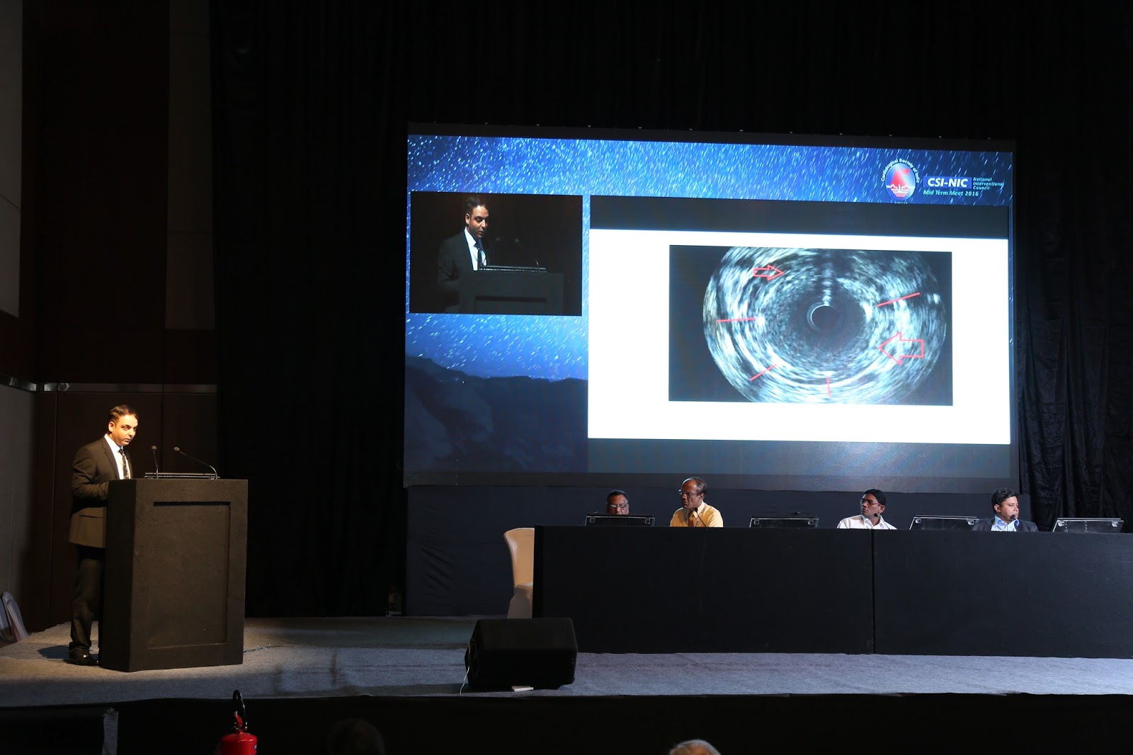
Coronary Calcium burden's detection by IVUS vs. angiography ( Acute stent fracture in Left main during primary PCI, Presentation in #NIC2016)
The extent of coronary artery calcium strongly correlates with the degree of atherosclerosis and, therefore, with the rate of future cardiac events. Coronary angiography has lowto-moderate sensitivity compared with grayscale intravascular ultrasound (IVUS) or optical coherence tomography (OCT), the gold standard for coronary calcium detection; but coronary angiography has a
relatively high positive predictive value.
relatively high positive predictive value.
The prevalence of severe calcium defined as superficial in nature with greater than 180 degree arc( which is detected by IVUS) , is estimated to present itself in 12% of cases using angiographic imaging. When intravascular ultrasound (IVUS) guidance is used, it is seen in approximately 26%of cases.
Presentation of Dr.Nabil Paktin Cardiology Online


Subscribe to:
Posts (Atom)