Tuesday, December 30, 2014

WATCHMAN Left Atrial Appendage Closure (LAAC) Therapy for AF Patients !

Stroke prevention in patients with nonvalvular atrial fibrillation (NVAF) has been the focus of substantial clinical investigation related to the increasing frequency of this arrhythmia with the aging population, the well-documented relationship between increasing age and increased stroke, and the particularly major morbidity/mortality from cardioembolic stroke The  widespread application of anticoagulant therapy, initially with warfarin, which has been proven superior to aspirin for stroke prevention . Multiple problems with warfarin, however, have been identified, including bleeding, contraindications to its application, patient compliance, and the need for routine monitoring .Thus, it is estimated that anticoagulation is not currently used in up to 50% of  eligible AF patients, which led to the development of new oral anticoagulants (NOACs), whose efficacy have been established in randomized clinical trials. Traditional treatment strategies have relied on chronic anticoagulation, either with warfarin or the newer anticoagulant agents,Anticoagulants like warfarin are typically the desired approach, but patients carry substantial risk of internal bleeding, either spontaneously or with minor trauma.  . Growing information regarding the central role of left atrial appendage (LAA) thrombus has led to mechanical approaches for stroke prevention in this setting.


 In the PROTECT AF (Watchman Left Atrial Appendage Closure Technology for Embolic Protection in
Patients With Atrial Fibrillation) trial that evaluated patients with nonvalvular atrial fibrillation (NVAF), left atrial appendage (LAA) occlusion was noninferior to warfarin for stroke prevention, but a periprocedural safety hazard was identified.

The most recent  PREVAIL trial stated ,  LAA occlusion was noninferior to warfarin for ischemic stroke prevention or SE >7 days’ post-procedure. Although noninferiority was not achieved for overall efficacy, event rates were low and numerically comparable in both arms. Procedural safety has significantly improved. This trial provides additional data that LAA occlusion is a reasonable alternative to warfarin therapy for stroke prevention in patients with NVAF who do not have an absolute contraindication to short-term warfarin therapy.


Sunday, December 21, 2014

Where adenosine can effect ?according to SVTs classification!

Adenosine,in addition to terminating supraventricular tachycardia involving the atrioventricular (AV) node, may have antiarrhythmic effects on atrial tachycardia but it is not effective for Atrial flutter and fibrillation.
The electrophysiological effects of adenosine on supraventricular tissue include shortening of action potential duration in atrial myocytes mediated by the potassium current, IKAChAdO; shortening of action potential duration and hyperpolarization in sinus node cells;and anti- adrenergic electrophysiological effects resulting from inhibition of adenylylcyclase.
In addition to its well known effects on AV nodal reentry and AV reciprocating tachycardia,as well as its effects on ventricular tachycardia due to triggered activity,adenosine is useful as a diagnostic probe for suspected sinus node reentrantt achycardia.
Furthermore,because adenosine has no effect on reentrant atrial tachycardia but instead transiently suppresses automatic atrial tachycardia and terminates atrial tachycardia presumed due to triggered activity,adenosine may be useful in differentiating between these arrhythmias.
Responsiveness of triggered atrial tachycardia to adenosine may account for some instances where adenosine has been previously thought to terminate intra-atrial reentrant tachycardia.

Usefulness of the Electrocardiogram in Predicting the Occlusion Site in Acute Anterior Myocardial Infarction with Isolated Disease of the Left Anterior

ST segment elevation in lead V1≥3 mm measured at 80 ms from the J point is associated in a statistically significant manner with a lesion proximal to the dominant septal artery, with 100% specificity( P=0.01).


Sunday, December 14, 2014

Are beta-blockers contraindicated for Asthma or COPD with heart failure patients?

There are 3 types of β receptors. β1-Adrenoceptors are situated in the cardiac sarcolemma. If activated, they lead to an increase in the rate and force of myocardial contraction (positive inotropic effect) by opening the calcium channels. On the other hand, β2-Adrenoceptors are found mainly in bronchial and vascular smooth muscles. If activated, they cause broncho- and vaso-dilatation. There are, however, sizable populations of β2-Adrenoceptors in the myocardium, of about 20%–25%, which leads to the cardiac effects of any β2-Adrenoceptors stimulation. There is a relative up-regulation of these receptors to about 50% in heart failure. The role of β3-Adrenoceptors in the heart is not yet fully identified and accepted .
Beta-blockers are classified into three generations. The first generation agents (such as Propranolol, Sotalol, Timolol, and Nadolol), are nonselective and block β1 and β2 receptors. Blocking β1-receptors affects the heart rate, conduction and contractility, while blocking β2-receptors, tends to cause smooth muscle contraction, therefore, bronchospasm in predisposed individuals. The second-generation agents or the cardioselective agents (such as Atenolol, Bisoprolol, Celiprolol, and Metoprolol) block β1-receptors in low doses but are capable of blocking β2-receptors in higher doses. This selective mode of action makes the use of these agents more suitable in patients with chronic lung disease or those with insulin-requiring diabetes mellitus. Cardioselectivity varies between agents with the Bisoprolol among the most selective. The third generation agents have vasodilatory properties. There action is either selective (Nebivolol) or nonselective (Carvidolol and Labetolol). The vasodilatory properties are mediated either by nitric oxide release as for Nebivolol or Carvidolol  or by added alpha-adrenergic blockade as in Labetolol and Carvidolol. A third vasodilatory mechanism, as in Pindolol and Acebutolol, acts via β2-intrinsic sympathomimetic activity (ISA).
 These beta-blockers therefore have the capacity to stimulate as well as to block adrenergic receptors and tend to cause less bradycardia than the other beta-blockers and may cause less coldness of the extremities.
Because of the bradyarrhythmic and hypotensive effects of beta-blockers, the major heart failure trials excluded patients with a heart rate of <50 to 68 beats per minute (BPm) or systolic blood pressure <80 to 100 mm hg (the ranges  cited reflect the variation in cut points from one study to another) and in clinical practice, physicians often withhold beta-blocker therapy from heart failure patients who also have chronic obstructive pulmonary disease (coPd) or  asthma, hypotension, or metabolic risk factors for diabetes.
Some avoid prescribing beta-blockers because they believe  that the drugs adversely affect patients’ quality of life, despite evidence to the contrary.In all these cases, there is  little justification for doing so.
Although beta-blockers can worsen and precipitate bronchospasm, recent evidence suggests that patients with coPd and asthma can tolerate them.In fact, there is reason to believe that bronchospasm is aggravated by excessive stimulation and sensitization of the beta-2 receptors, and that blocking them may even be  of therapeutic value. nonetheless, the danger of worsening bronchospasm with a nonselective beta-blocker such as  carvedilol remains—particularly for patients with asthma, who tend to have a higher degree of bronchial sensitivity and  reactivity. So, while beta-blockers are not contraindicated for patients with coPd, their use in this patient population  requires caution.


Thursday, December 11, 2014

Palpitations after Dinner (Non-Cardiac Cause of Palpitation)


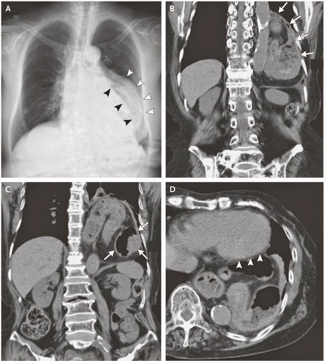
A 76-year-old woman with rheumatoid arthritis, diabetes mellitus, and hypertension presented with a 1-month history of palpitations that occurred only after she had eaten dinner. The sensation was felt at the center of the chest and lasted for 10 to 15 minutes after the meal. An electrocardiogram was unremarkable. A chest radiograph (Panel A) showed a mediastinal shadow (white arrowheads) lateral to the left heart border (black arrowheads). Computed tomography of the chest revealed a left diaphragmatic hernia (Panel B), with the stomach positioned in the thorax (Panel C, coronal view), abutting the left ventricle (Panel D, axial view, arrowheads). The stomach was visibly twisted, a finding consistent with a gastric volvulus. Gastric endoscopy revealed a volvulus, with twisting of the mucosa. After surgical repair of the hernia and volvulus, the palpitations resolved, and at follow-up more than 1 year after surgery, the patient remained free of symptoms.


Reference:
Kazutaka Kurokohchi, M.D., Ph.D., and Osamu Imataki, M.D.
N Engl J Med 2014; 371:2320

Tuesday, December 9, 2014

Intrarenal effects of angiotensin-converting (ACE) inhibitors and angiotensin receptor blockers (ARBs)

The manner in which renal function changes when an ACE inhibitor is started depends on the treatment circumstances. In most instances there is little to no change in serum creatinine value (or the level of renal function) when an ACE inhibitor is started. This is typically the case when hypertension alone is being treated. Occasionally, renal function declines (by 10% to 20%) shortly after an ACE inhibitor is  started, a process that arises from a resetting of renal hemodynamics and is fully reversible when the drug is discontinued.
This pattern is observed in patients with underlying renal disease. The magnitude of this early drop in renal function may, in fact, identify those patients likely to benefit most from ACE inhibitor therapy.
Concern engendered by this change in renal function is unfounded. A more substantial drop in renal function is occasionally seen in patients who are being treated with an ACE inhibitor. This occurs most commonly in patients who are either volume contracted and/or have bilateral large or small artery disease in the renal vascular bed. Correction of any volume deficit typically returns renal function to baseline values. In the
instance of renal arterial disease, reducing the dosage of an ACEinhibitor (or, if necessary, discontinuing it) will restore renal function to its baseline level. The change in renal function with ARBs given to patients with or without renal disease appears to differ little from that seen with ACE inhibitors.
In the untreated state efferent arteriolar tone is presumably increased and glomerular hyperfiltration exists (left panel). The change in the diameter of the efferent arteriole is greater with ACE inhibition, reflecting the combined effect of increased bradykinin and decreased angiotensin II concentrations on efferent arteriolar tone (middle). A proposed consequence of this change is a somewhat greater drop in the glomerular filtration rate with an ACE inhibitor. In the case of ARBs the absence of a direct effect on bradykinin limits any decrease in efferent arteriolar tone to what might occur with a reduction in angiotensin II effect (right). Although this scenario has been demonstrated experimentally, confirming evidence of this process is still not available in humans.


Sunday, December 7, 2014

Assessment of MR Severity ( Color Flow Doppler )

 The colour flow area of the regurgitant jet is not recommended to quantify the severity of MR. The colour flow imaging should only be used for detecting MR. A more quantitative approach is required when more than a small central MR jet is observed.

Wednesday, December 3, 2014

What is Slow Coronary Flow (SCF) Phenomenon?

Cardiologists are familiar with the phenomenon of slow progression of angiographic contrast in the coronary arteries in the absence of stenosis in the epicardial vessels in some patients presenting with chest pain. The coronary slow flow phenomenon (CSFP), first described in 1972, remains scantily studied. This phenomenon should be distinguished from occurrence of slow flow in the context of coronary reperfusion therapy such as angioplasty or thrombolysis that is associated with different pathophysiological and clinical implications. Similarly, coronary slow flow associated with coronary artery spasm, coronary artery ectasia (CAE), myocardial dysfunction, valvular heart disease and certain connective tissue disorders involving coronary microvasculature is easy to understand.CSFP may occassionly also result from inadvertent air-embolism during angiography or may be due to an overlooked ostial lesion. However, it is not certain whether CSFP in the absence of any of these known causes represents merely an angiographic curiosity or has special physiologic or therapeutic implications. In this editorial we focus on the current knowledge regarding CSFP manifesting in the absence of any known etiology.


In slow Coronary Flow (SCF)  phenomenon  there is a delayed opacification opacification  of epicardial  arteries in the absence of occlusive disease.Although the pathogenesis of this syndrome is controversial, several several  studies have suggested that  it is mainly caused by endothelial microvascular dysfunction.There is also a correlation between SCF and abnormal hemorheologic  parameter.
 Hematocrit and Current Smoking (15% vs. 39%)were the only variables that were found to be were the only variables that were found to be significant when comparing the two groups of significant when comparing the two groups of patients.
In patients with normal coronary arteries coronary blood flow velocity is highly correlated
between different blood vessels and t the different measuring techniques. different measuring techniques.  Slow flow is highly correlated to Hematocrit Current Smoking,blood viscosity impairs 
blood flow.
Smoking may involve the induction of endothelial dysfunction, moreover, smoking is known to elevate hematocrit level and perhaps, by doing so, it increases blood viscosity which slower the blood flow even more.

Sunday, November 30, 2014

SLOW ("POOR") R WAVE PROGRESSION

The R wave height normally becomes progressively taller from leads V1 through V6. In V1 to V3, there is an R wave of low amplitude and an S wave that is larger (R/S <1); between leads V3 to V4, there is a transition to an R wave that has a greater amplitude than the S wave (R/S >1). When the R wave height does not become progressively taller from leads V1 to V3 or V4, or even remains at low amplitude across the entire precordium, slow or poor R wave progression (PRWP) is present . Several sets of criteria have been proposed to define PRWP more precisely .

There are many etiologies for PRWP:
●Lead placement
●Anteroseptal or anterior wall myocardial infarction
●Left ventricular hypertrophy
●Left anterior fascicular block
●Left bundle branch block
●Infiltrative or dilated cardiomyopathy
●Wolff-Parkinson-White patterns due to right-sided or antero-septal bypass tracts
●Chronic lung disease
●Physiologic late transition or clockwise rotation of the heart
Poor R wave progression may be more frequently seen in females. Because there are so many different causes of PRWP, this finding alone is not useful in identifying patients with a prior anterior myocardial infarction.

Flying Defibrillator

Let’s Use Drones for Good Purpose, Let’s Use Drones to save Lives.
The traffic overcrowd cannot be the  reason of delay for cardiac arrested ones any more .
This improves current emergency infrastructure services to save the life, the ultra-fast response system moving 100 kilometers per hour which increasing the survival chance from 8% to 80% .
This can bring the average ambulance response time from 10 minutes to one .

This is equipped with camera and GPS navigation system , this will be capable of carrying oxygen mask and some injections .

Monday, November 24, 2014

Myocardial bridging:Normal , Abnormal or both !

In 5-12% of patients coronary artery Courses intramyocardial instead of coursing epicardially , this congenital coronary anomaly called Myocardial Bridging..Muscle overlying the intramyocardial segment of an epicardial coronary artery, first mentioned by Reyman in 1737, is termed a myocardial bridge, and the artery coursing within the myocardium is called a tunneled artery.


It is characterized by systolic compression of the tunneled segment, which remains clinically silent in the vast majority of cases.Coronary atherosclerosis in association with myocardial bridging has primarily been studied in the LAD. The segment proximal to the bridge frequently shows atherosclerotic plaque formation, although the tunneled segment is typically spare.
Hemodynamic forces may explain atherosclerotic plaque formation at the entrance to the tunneled segment. There, the endothelium is flat, polygonal, and polymorph, indicating low shear, whereas in the tunneled segment, the endothelium has a helical, spindle-shaped orientation along the course of the segment as a sign of laminar flow and high shear. Low shear stress may induce the release of endothelial vasoactive agents such as endothelial nitric oxide synthase (eNOS), endothelin-1 (ET-1), and angiotensin-converting enzyme (ACE). Their levels were significantly higher in proximal and distal segments compared with the tunneled segment. Thus, low shear stress may contribute to atherosclerotic plaque formation proximal to the bridge, whereas high shear stress may have a protective role within the tunneled segment. In addition, an increase in local wall tension and stretch may induce endothelial injury and plaque fissuring with subsequent thrombus formation in the proximal segment,which is supported by autopsy and clinical observations.
For further reading Please Click Read More ...

Sunday, November 23, 2014

Blue Toe Syndrome ( Cholesterol Embolization Syndrome)

Blue toe syndrome is characterised by tissue ischaemia secondary to cholesterol crystal or atherothrombotic embolisation. It leads to the occlusion of small vessels. Cyanosis of the digits may have several etiologies ranging from trauma to connective tissue disease, however the most common cause of blue toe syndrome is atheroembolic disease or aneurysm. Embolisation occurs typically following an ulcerated atherosclerotic plaque or aneurysms located in the aorto-iliac-femoral arterial system. Embolisation can occur spontaneously or due to a variety of causes. Most often, microembolisation appears in elderly men who have undergone an angiographic procedure or vascular surgery or even anticoagulant or thrombolytic treatment .
The differential diagnosis of blue toe syndrome includes Raynaud’s (especially secondary) phenomenon. In Raynaud’s syndrome ishaemic lesions are usually more diffuse, larger areas of distal parts of different fingers (rarely toes) are involved and are affected by vasospastic disorders. The patients are usually younger and without any known atherosclerotic disease. In blue toe syndrome skin lesions are usually restricted and related to the occluded artery, fingers are rarely affected as opposed to toes and vasospasm is usually absent. Painful red, purple, blue or black toes can also be seen in patients with thrombocythemia associated with polycythemia vera or in essential thrombocythemia (3). In these patients, ischemic lesions of fingers or toes are caused by arteriolar inflammation and the thrombosis of microvessels that appear as a consequence.

 The following 6 key elements are required for the development of cholesterol embolization syndrome:
  1. Presence of a plaque in a proximal, large-caliber artery (such as the internal carotid artery, the iliac arteries, or the aorta)
  2. Plaque rupture (spontaneous, traumatic, or iatrogenic)
  3. Embolization of plaque debris (containing cholesterol crystals, platelets, fibrin, and calcified detritus)
  4. Lodging of the emboli in small to medium arteries with a diameter of 100 to 200 μm, leading to mechanical occlusion
  5. Foreign-body inflammatory response to cholesterol emboli
  6. End-organ damage due to a combined effect of mechanical plugging and inflammation.

Saturday, November 22, 2014

Definitions of PCI Success

Percutaneous coronary intervention (PCI) success may be defined by angiographic, procedural, and clinical criteria. 

1•Angiographic success in a stented artery is a minimum stenosis diameter reduction to <20% .

2-Procedural Success
A successful PCI : should achieve angiographic success without in-hospital major clinical complications (e.g., death, myocardial infarction [MI], emergency coronary artery bypass surgery) during hospitalization. MI is often defined as the development of Q waves in addition to elevation of troponins three times the upper limit of the laboratory's normal value. Cardiac troponin T and I as measurements of myocardial necrosis are more sensitive and specific than CK-MB. Enzyme elevation in the absence of new Q waves is counted as MI, peri-procedural. There is no consensus on what level of troponin alone is clinically important enough to change major management following the interventional procedure.

3-Clinical Success •A clinically successful PCI is ananatomical and procedural success with relief of signs and/or symptoms of myocardial ischemia after recovery from the procedure. The long-term clinical success requires that the patient have persistent relief of signs and symptoms of myocardial ischemia for more than 6 months. Restenosis is the principal cause of lack of long-term clinical success when short-term clinical success has been achieved.


Wednesday, November 5, 2014

ACE Inhibitors Side Effect Mnemonic (CAPTOPRIL)


News and Views :Antibiotic Cotrimoxazole Combined With ACE Inhibitor or ARB Could Raise Risk of Sudden Death

October 31, 2014
TORONTO, ON — The risk of sudden death went up by more than a third in older patients taking ACE inhibitors or angiotensin-receptor blockers (ARBs) who were also put on the antibacterial agent cotrimoxazole, compared with those who were instead given amoxicillin, in a case-control study reported this week
. The finding was independent of comorbidities, other medications, recent procedures, and other potential influencers of sudden-death risk, according to the authors.The elevated risk with cotrimoxazole, a combination of sulfamethoxazole and trimethoprim widely used for decades, was likely caused by its capacity for raising serum potassium, which became fatal on top of other medications known for causing hyperkalemia, speculate Dr Michael Fralick (University of Toronto, ON) and colleagues in their report, published October 30, 2014 in the BMJ. The same group had previously observed that combining cotrimoxazole with ACE inhibitors or ARBs similarly drove up the risk of hospitalization due to hyperkalemia.
"In patients who are on ACE inhibitors or ARBs, who are by definition at risk for hyperkalemia, the safest thing to do would be to use an antibiotic" other than cotrimoxazole, senior author Dr David N Juurlink (Institute for Clinical Evaluative Sciences and the University of Toronto, ON) told heartwire . "But that's not always going to be practical. Alternate strategies could be to use a lower dose or a shorter duration of the drug. Or, when you have to give trimethoprim-based antibiotics to somebody who's on an ACE inhibitor or an ARB, at a minimum being mindful of the potential for serious and even life-threatening hyperkalemia. That alone would go a long way toward reducing the dangers of this interaction."
That goes especially for some susceptible patient groups. "Patients with type 2 diabetes are sitting ducks for this," Juurlink said. "They have a tendency for hyperkalemia independent of anything else, because of what the diabetes does to their kidney function." And patients with heart failure may be on potassium-sparing spironolactone as well as ACE inhibitors or ARBs. "Someone who has reduced left ventricular function and has diabetes—there are millions of people like that in North America."
For further reading Please click Read More 

Wednesday, October 15, 2014

News and Views : Increased Mortality Associated With Digoxin in Contemporary Patients With Atrial Fibrillation Findings From the TREAT-AF Study


News and Views ; Atrial fibrillation associated with ivabradine treatment: meta-analysis of randomised controlled trials

ABSTRACT
Objective :To quantify any risk of atrial fibrillation (AF) associated with ivabradine treatment by meta-analysis of clinical trial data.
Methods Medline, Embase, Web of Knowledge and the Cochrane central register of controlled trials were searched for double-blinded randomised controlled trials of ivabradine with a minimum follow-up period of 4 weeks. For studies where AF data were unpublished, safety data were obtained from the European Medicines
Agency (EMeA) website and personal communications. Studies were appraised for risk of bias using components recommended by the Cochrane Collaboration. Metaanalyses were performed of relative risk of AF and absolute risk difference of AF per year of treatment. The main outcome measure was incident AF during the follow-up period.
Results: AF data were available from 11 studies: one from the published report, six from the EMeA and four from personal communications. Ivabradine treatment was associated with a relative risk of AF of 1.15 (95% CI
1.07 to 1.24, p=0.0027) among 21 571 patients in the meta-analysis. From this we estimated that the number needed to harm for ivabradine would be 208 (95% CI 122 to 667) per year of treatment.
Conclusions:
AF is a substantially more common side effect of ivabradine treatment than one patient in 10 000, the risk presently reported in the product literature. The incidence of AF has not routinely been reported in clinical trials of ivabradine. Ivabradine treatment is associated with a 15% increase in the RR of AF. We estimate that 208 patient-years of treatment with ivabradine would be required to cause one new case of AF.
Martin RIR, et al. Heart 2014;100:1506–1510. doi:10.1136/heartjnl-2014-305482
BMJ

Wednesday, September 17, 2014

From ICCU to ICU bedside Echocardiography !

However, Bedside transthoracic echocardiography (TTE) provides rapid and noninvasive hemodynamic assessment of critically ill patients but is limited by the immediate availability of experienced sonographer .Noncardiologists   are  able to estimate LV function with reasonable accuracy using a hand-held unit in the ICU ,but,  indeed, noncardiologists  often fail to identify important cardiac abnormalities such as valvular disease and regional wall abnormalities, important cardiac causes of hemodynamic compromise such as cor pulmonale, acute valvular abnormalities, and intracardiac shunts as well as very complicated cases rather than LV function determination only  .
Its training should be mandatory  for all intensivists and anesthesiologist  because it is of particular importance .

For example,respiratory variation in inferior vena cava diameter may help predict the likelihood of fluid responsiveness, and measurement of flow across the aortic valve can be used to estimate cardiac output and calculate systemic vascular resistance.
 Inotropic therapy might be withheld from patients with hemodynamic impairment resulting from decreased LV function if the TTE was misinterpreted as normal. Conversely, a patient with normal LV function whose hypotension was due to hypovolemia or vasodilation might be inappropriately treated with an inotrope if LV function was incorrectly deemed to be abnormal. in a daily basis , the most common error was failure to recognize a decrease in LV function rather than misinterpreting normal LV function as abnormal.  

Common variations in papillary muscle anatomy in HCM


Monday, September 15, 2014

Choice of Drugs for Cardiac Arrhythmias

Symptomatic tachycardias and premature beats may be treated with a variety of antiarrhythmic drugs. These may be given intravenously in an emergency situation or orally for long-term treatment. These drugs either suppress the abnormal firing of pacemaker tissue or depress the transmission of impulses in tissues that either conduct too rapidly or participate in reentry.

The relative simplicity of antiarrhythmic drug therapy must be balanced against two disadvantages. One is that the drugs must be taken daily and indefinitely. The other is the risk of side effects. While side effects are a risk of all medication, those associated with antiarrhythmic drugs can be very hard to manage. They include proarrhythmia, the more-frequent occurrence of preexisting arrhythmias or the appearance of new arrhythmias as bad as or worse than those being treated.


Saturday, September 13, 2014

Prominent Papillary Muscle or Papillary Fibroelastoma, Apically displaced papillary muscles,Solitary Papillary Muscle Hypertrophy

A 55 years old female , brought to the Echo Lab for perioperative assessment of non-cardiac surgery of the heart . She was known patient of hypertension , DM type 2 with normal physical examination . During echocardiography we found a mass in LV cavity ECG was normal with no axis deviation , and hypertrophy pattern . There was Cardiologist to cardiologist difference of opension about below answers of diagnosis of the following echocardiogram :
1- Prominent Papillary muscle .
2- Papillary Fibroblastoma 
3- Apically displaced anterolateral papillary muscle .
4-  Solitary Papillary Muscle Hypertrophy.

The apically displaced anterolateral papillary muscle was definedwhen the base of the papillary muscle was located at the apical one-third of the left ventricle.
An abnormal insertion of the papillary muscle was noted in some patients with ADPM. Some patients had papillary muscle inserted into the base of a mitral leaflet or directly into the LVOT.


Apical hypertrophic cardiomyopathy (ApHCM) is a subtype ofmhypertrophic cardiomyopathy (HCM), which is more frequently mfound in Asians than in Caucasians.Although it has been demonstrated that ApHCM is generally benign, cardiovascular complications are not uncommon and therefore, correct diagnosis of mthis entity is important. Twelve-lead electrocardiograms (ECGs) of ApHCM are characterized by a giant negative T (GNT) wave in precordial leads. Moreover, the depth of the GNT wave has been reported to be associated with the severity of apical thickening or with the ratio of apical-to-basal myocardial thickness. Therefore, in the presence of a GNT wave, a diagnosis of ApHCM has often been made in patients with apical hypertrophy, especially when the GNT wave cannot be explained otherwiseThe phenomenon of solitary papillary muscle hypertrophy is rare with only 2 references in the literature. Furthermore, giant negative T and U waves are 2 common electrocardiographic phenomena in hypertrophic cardiomyopathy and have been attributed to hypertrophy of the posterior papillary muscle. Solitary hypertrophy of the anterior papillary muscle might be a new echo-electrocardiographic syndrome.
Electrocardiogram and its images of apical hypertrophy. (A) T wave negativity more prominent in the mid-precordial than in the lateral precordial leads represent classic electrocardiographic finding in apical hypertrophy. (B) Representative echocardiography images of the classic pattern of apical hypertrophic cardiomyopathy showing an ‘ace of spade’ shape during diastole. (C) However, it is not unusual to encounter an atypical pattern of hypertrophy showing more prominent hypertrophy at the apical lateral than at the apical septal segments.
Various patterns of anterolateral papillary muscle location in the apical four-chamber view. (A) Normally located papillary muscle . (B) Apically displaced papillary muscle . (C) Variation in the morphology of papillary muscle head attached to the left ventricular (LV) . (D) Apically attached accessory papillary muscle. (E) The papillary muscle inserted into the base of a mitral valve leaflet . (F) The papillary muscle inserted into the left ventricular outflow tract . An apicoseptal segment may also show certain degree of hypertrophy, but for the clarity of presentation, this finding is not represented in above fig.


Anothoer distinc feature of papillary muscle is Papillary fibroelastoma, or papilloma, is a benign intracardiac tumor with a characteristic appearance on echocardiograms. It is usually small (average size, <15 mm) and has a characteristic stippled edge, with a shimmering appearance at the tumor-blood interface. There are f ingerlike projections consistent with the fronds that are described pathologically as a “sea anemone.” The most frequent location is the aortic valve on either the aortic or ventricular surface, followed by either the atrial or ventricular surface of the mitral valve.138 Papillomas can be found in any chamber or on any surface and are usually single, but multiple tumors occur in approximately 10% of patients.
Solitary Papillary Muscle Hypertrophy: Transthoracic, two-dimensional echocardiography revealed isolated hypertrophy of the anterolateral papillary muscle, with an otherwise normal left ventricle with no hypertrophy in any other segment( image below) .  

The answer is A. Prominent Papillary Muscle . 


Wednesday, September 10, 2014

Dilated Cardiomyopathy and Myocardial Infarction

  •  Cardiac magnetic resonance (CMR) is the current golden standard for accurate assessment of ventricular volumes and global and regional function, thus providing information on prognosis.
    • Extracellular accumulation of gadolinium, imaged at 10 to 30 minutes after injection, identifies areas of myocardial fibrosis commonly seen in cardiomyopathies, generally subendocardial or transmural in a coronary distribution in the case of ischemic cardiomyopathy.
    • Transmural extent of LGE can predict recovery of myocardial function after revascularization in patients with coronary artery disease.
    • Negative stress CMR with either adenosine or dobutamine stressors has excellent negative predictive value for subsequent coronary events.
    • Noncoronary segmental distribution and midwall or epicardial involvement of LGE are atypical of myocardial infarction. This allows discrimination between ischemic and nonischemic etiologies of cardiomyopathy, which can assist in clinical decision making.
    • Takotsubo cardiomyopathy is characterized by left ventricular apical dilation and regional hypocontraction, absence of first-pass perfusion abnormality, and minimal or absent concomitant subendocardial LGE.
    • Because of its higher spatial and temporal resolution, CMR can offer an improved assessment for the presence and extend of ventricular noncompaction, compared with echocardiography.
    • CMR can accurately quantify severity of valvular regurgitation and stenosis using phase contrast imaging. In the same study CMR can assess the physiologic impact of the valvular heart disease by providing quantitation of large vessel morphology and ventricular volumes and functions.
    • CMR is the most sensitive technique for detecting clinically unrecognized small subendocardial myocardial infarctions that do not result in wall motion abnormality or ECG changes, thereby identifying patients at risk of cardiac events.
    • CMR infarct imaging can provide direct information on the amount of irreversibly injured myocardium and viable myocardium, including the extent of periinfarct zone and microvascular obstruction, which are strong predictors of cardiovascular outcome in patients after myocardial infarction (MI).
     Case: A 54-year-old woman without significant risk factors for coronary artery disease presented to our services complaining of a 4-week history of exertional dyspnea (NYHA class III) and pedal edema. The patient did not have any history of alcoholism, family history of heart disease, or sudden cardiac death. ECG showed sinus rhythm and a left bundle branch block pattern of unknown duration. Transthoracic echocardiography showed a dilated left ventricle and severe global hypokinesis with reduced ejection fraction of 25%. A coronary angiography was performed that revealed no significant coronary stenosis. CMR was performed .

  • For More Reading Please Click Read More !