Sunday, April 24, 2016

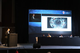
Coronary Calcium burden's detection by IVUS vs. angiography ( Acute stent fracture in Left main during primary PCI, Presentation in ‪#‎NIC2016‬)

The extent of coronary artery calcium strongly correlates with the degree of atherosclerosis and, therefore, with the rate of future cardiac events. Coronary angiography has lowto-moderate sensitivity compared with grayscale intravascular ultrasound (IVUS) or optical coherence tomography (OCT), the gold standard for coronary calcium detection; but coronary angiography has a
relatively high positive predictive value.
The prevalence of severe calcium defined as superficial in nature with greater than 180 degree arc( which is detected by IVUS) , is estimated to present itself in 12% of cases using angiographic imaging. When intravascular ultrasound (IVUS) guidance is used, it is seen in approximately 26%of cases.

Friday, April 1, 2016

Perspective on the stethoscope at 200 years old

200 years ago, in 1816, French physician René Laennec rolled a sheet of paper into a tube to auscultate the chest of a young female patient with suspected tuberculosis. Laennec went on to make his first stethoscope from two pieces of hollowed wood: one was placed against the physician's ear; the other, with a funnel-shaped cone at the end, was placed on the patient's chest. Little did Laennec realise the scientific maelstrom that would greet this new diagnostic instrument, and indeed the parallels that could be drawn to the introduction of handheld ultrasound into clinical practice some 200 years later.


Why there will be Right to left shunting in RV infarction ?

For better resolution of this image, please click on it and maximize it . 

Giant Aneurysm of the Right Coronary Artery: a Rare Cause of Atrial Fibrillation

A 46-year old patient, with no cardiovascular risk factors, sought treatment for frequent episodes of irregular heartbeat and stabbing chest pain, accompanied by dyspnea and nausea at rest, which was directly related with lying on his right side. In one of these episodes, we documented atrial fibrillation with a rapid ventricular response, which spontaneously revertedto sinus rhythm. The only relevant background in the patient's medical history was hypothyroidism secondary to a thyroidectomy resulting from multi-nodular goitre. The patient was euthyroid, and received treatment with levothyroxine. A trans-thoracic echocardiogram revealed a large heteroechoic cyst-like mass behind the right atrium that was severely depressing this structure along with the left atrium, to a lesser degree (Figure 1). A computed tomography revealed a giant aneurysm of the right coronary artery, 8×8.8 cm in size, with a heterogeneous density. It emerged immediately adjacent to the root of the right coronary artery and displaced and compressed both atria (Figure 2). After confirming the diagnosis by coronary angiography (Figure 3), the patient underwent surgical exclusion of the aneurysm and plication of the atrium (right atriotomy). The patient has had a favorable postoperative evolution, with no relevant complications.


Source : Cardiologia