Tuesday, December 30, 2014

WATCHMAN Left Atrial Appendage Closure (LAAC) Therapy for AF Patients !

Stroke prevention in patients with nonvalvular atrial fibrillation (NVAF) has been the focus of substantial clinical investigation related to the increasing frequency of this arrhythmia with the aging population, the well-documented relationship between increasing age and increased stroke, and the particularly major morbidity/mortality from cardioembolic stroke The  widespread application of anticoagulant therapy, initially with warfarin, which has been proven superior to aspirin for stroke prevention . Multiple problems with warfarin, however, have been identified, including bleeding, contraindications to its application, patient compliance, and the need for routine monitoring .Thus, it is estimated that anticoagulation is not currently used in up to 50% of  eligible AF patients, which led to the development of new oral anticoagulants (NOACs), whose efficacy have been established in randomized clinical trials. Traditional treatment strategies have relied on chronic anticoagulation, either with warfarin or the newer anticoagulant agents,Anticoagulants like warfarin are typically the desired approach, but patients carry substantial risk of internal bleeding, either spontaneously or with minor trauma.  . Growing information regarding the central role of left atrial appendage (LAA) thrombus has led to mechanical approaches for stroke prevention in this setting.


 In the PROTECT AF (Watchman Left Atrial Appendage Closure Technology for Embolic Protection in
Patients With Atrial Fibrillation) trial that evaluated patients with nonvalvular atrial fibrillation (NVAF), left atrial appendage (LAA) occlusion was noninferior to warfarin for stroke prevention, but a periprocedural safety hazard was identified.

The most recent  PREVAIL trial stated ,  LAA occlusion was noninferior to warfarin for ischemic stroke prevention or SE >7 days’ post-procedure. Although noninferiority was not achieved for overall efficacy, event rates were low and numerically comparable in both arms. Procedural safety has significantly improved. This trial provides additional data that LAA occlusion is a reasonable alternative to warfarin therapy for stroke prevention in patients with NVAF who do not have an absolute contraindication to short-term warfarin therapy.


Sunday, December 21, 2014

Where adenosine can effect ?according to SVTs classification!

Adenosine,in addition to terminating supraventricular tachycardia involving the atrioventricular (AV) node, may have antiarrhythmic effects on atrial tachycardia but it is not effective for Atrial flutter and fibrillation.
The electrophysiological effects of adenosine on supraventricular tissue include shortening of action potential duration in atrial myocytes mediated by the potassium current, IKAChAdO; shortening of action potential duration and hyperpolarization in sinus node cells;and anti- adrenergic electrophysiological effects resulting from inhibition of adenylylcyclase.
In addition to its well known effects on AV nodal reentry and AV reciprocating tachycardia,as well as its effects on ventricular tachycardia due to triggered activity,adenosine is useful as a diagnostic probe for suspected sinus node reentrantt achycardia.
Furthermore,because adenosine has no effect on reentrant atrial tachycardia but instead transiently suppresses automatic atrial tachycardia and terminates atrial tachycardia presumed due to triggered activity,adenosine may be useful in differentiating between these arrhythmias.
Responsiveness of triggered atrial tachycardia to adenosine may account for some instances where adenosine has been previously thought to terminate intra-atrial reentrant tachycardia.

Usefulness of the Electrocardiogram in Predicting the Occlusion Site in Acute Anterior Myocardial Infarction with Isolated Disease of the Left Anterior

ST segment elevation in lead V1≥3 mm measured at 80 ms from the J point is associated in a statistically significant manner with a lesion proximal to the dominant septal artery, with 100% specificity( P=0.01).


Sunday, December 14, 2014

Are beta-blockers contraindicated for Asthma or COPD with heart failure patients?

There are 3 types of β receptors. β1-Adrenoceptors are situated in the cardiac sarcolemma. If activated, they lead to an increase in the rate and force of myocardial contraction (positive inotropic effect) by opening the calcium channels. On the other hand, β2-Adrenoceptors are found mainly in bronchial and vascular smooth muscles. If activated, they cause broncho- and vaso-dilatation. There are, however, sizable populations of β2-Adrenoceptors in the myocardium, of about 20%–25%, which leads to the cardiac effects of any β2-Adrenoceptors stimulation. There is a relative up-regulation of these receptors to about 50% in heart failure. The role of β3-Adrenoceptors in the heart is not yet fully identified and accepted .
Beta-blockers are classified into three generations. The first generation agents (such as Propranolol, Sotalol, Timolol, and Nadolol), are nonselective and block β1 and β2 receptors. Blocking β1-receptors affects the heart rate, conduction and contractility, while blocking β2-receptors, tends to cause smooth muscle contraction, therefore, bronchospasm in predisposed individuals. The second-generation agents or the cardioselective agents (such as Atenolol, Bisoprolol, Celiprolol, and Metoprolol) block β1-receptors in low doses but are capable of blocking β2-receptors in higher doses. This selective mode of action makes the use of these agents more suitable in patients with chronic lung disease or those with insulin-requiring diabetes mellitus. Cardioselectivity varies between agents with the Bisoprolol among the most selective. The third generation agents have vasodilatory properties. There action is either selective (Nebivolol) or nonselective (Carvidolol and Labetolol). The vasodilatory properties are mediated either by nitric oxide release as for Nebivolol or Carvidolol  or by added alpha-adrenergic blockade as in Labetolol and Carvidolol. A third vasodilatory mechanism, as in Pindolol and Acebutolol, acts via β2-intrinsic sympathomimetic activity (ISA).
 These beta-blockers therefore have the capacity to stimulate as well as to block adrenergic receptors and tend to cause less bradycardia than the other beta-blockers and may cause less coldness of the extremities.
Because of the bradyarrhythmic and hypotensive effects of beta-blockers, the major heart failure trials excluded patients with a heart rate of <50 to 68 beats per minute (BPm) or systolic blood pressure <80 to 100 mm hg (the ranges  cited reflect the variation in cut points from one study to another) and in clinical practice, physicians often withhold beta-blocker therapy from heart failure patients who also have chronic obstructive pulmonary disease (coPd) or  asthma, hypotension, or metabolic risk factors for diabetes.
Some avoid prescribing beta-blockers because they believe  that the drugs adversely affect patients’ quality of life, despite evidence to the contrary.In all these cases, there is  little justification for doing so.
Although beta-blockers can worsen and precipitate bronchospasm, recent evidence suggests that patients with coPd and asthma can tolerate them.In fact, there is reason to believe that bronchospasm is aggravated by excessive stimulation and sensitization of the beta-2 receptors, and that blocking them may even be  of therapeutic value. nonetheless, the danger of worsening bronchospasm with a nonselective beta-blocker such as  carvedilol remains—particularly for patients with asthma, who tend to have a higher degree of bronchial sensitivity and  reactivity. So, while beta-blockers are not contraindicated for patients with coPd, their use in this patient population  requires caution.


Thursday, December 11, 2014

Palpitations after Dinner (Non-Cardiac Cause of Palpitation)


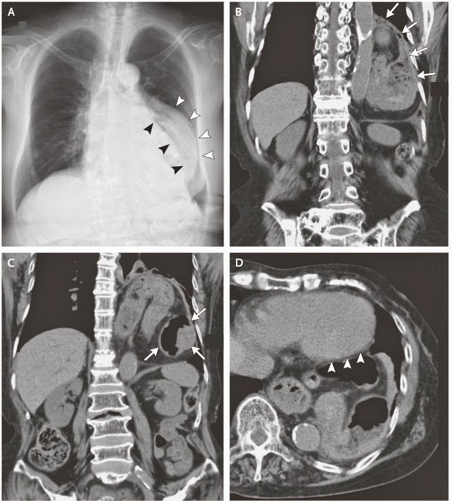
A 76-year-old woman with rheumatoid arthritis, diabetes mellitus, and hypertension presented with a 1-month history of palpitations that occurred only after she had eaten dinner. The sensation was felt at the center of the chest and lasted for 10 to 15 minutes after the meal. An electrocardiogram was unremarkable. A chest radiograph (Panel A) showed a mediastinal shadow (white arrowheads) lateral to the left heart border (black arrowheads). Computed tomography of the chest revealed a left diaphragmatic hernia (Panel B), with the stomach positioned in the thorax (Panel C, coronal view), abutting the left ventricle (Panel D, axial view, arrowheads). The stomach was visibly twisted, a finding consistent with a gastric volvulus. Gastric endoscopy revealed a volvulus, with twisting of the mucosa. After surgical repair of the hernia and volvulus, the palpitations resolved, and at follow-up more than 1 year after surgery, the patient remained free of symptoms.


Reference:
Kazutaka Kurokohchi, M.D., Ph.D., and Osamu Imataki, M.D.
N Engl J Med 2014; 371:2320

Tuesday, December 9, 2014

Intrarenal effects of angiotensin-converting (ACE) inhibitors and angiotensin receptor blockers (ARBs)

The manner in which renal function changes when an ACE inhibitor is started depends on the treatment circumstances. In most instances there is little to no change in serum creatinine value (or the level of renal function) when an ACE inhibitor is started. This is typically the case when hypertension alone is being treated. Occasionally, renal function declines (by 10% to 20%) shortly after an ACE inhibitor is  started, a process that arises from a resetting of renal hemodynamics and is fully reversible when the drug is discontinued.
This pattern is observed in patients with underlying renal disease. The magnitude of this early drop in renal function may, in fact, identify those patients likely to benefit most from ACE inhibitor therapy.
Concern engendered by this change in renal function is unfounded. A more substantial drop in renal function is occasionally seen in patients who are being treated with an ACE inhibitor. This occurs most commonly in patients who are either volume contracted and/or have bilateral large or small artery disease in the renal vascular bed. Correction of any volume deficit typically returns renal function to baseline values. In the
instance of renal arterial disease, reducing the dosage of an ACEinhibitor (or, if necessary, discontinuing it) will restore renal function to its baseline level. The change in renal function with ARBs given to patients with or without renal disease appears to differ little from that seen with ACE inhibitors.
In the untreated state efferent arteriolar tone is presumably increased and glomerular hyperfiltration exists (left panel). The change in the diameter of the efferent arteriole is greater with ACE inhibition, reflecting the combined effect of increased bradykinin and decreased angiotensin II concentrations on efferent arteriolar tone (middle). A proposed consequence of this change is a somewhat greater drop in the glomerular filtration rate with an ACE inhibitor. In the case of ARBs the absence of a direct effect on bradykinin limits any decrease in efferent arteriolar tone to what might occur with a reduction in angiotensin II effect (right). Although this scenario has been demonstrated experimentally, confirming evidence of this process is still not available in humans.


Sunday, December 7, 2014

Assessment of MR Severity ( Color Flow Doppler )

 The colour flow area of the regurgitant jet is not recommended to quantify the severity of MR. The colour flow imaging should only be used for detecting MR. A more quantitative approach is required when more than a small central MR jet is observed.

Wednesday, December 3, 2014

What is Slow Coronary Flow (SCF) Phenomenon?

Cardiologists are familiar with the phenomenon of slow progression of angiographic contrast in the coronary arteries in the absence of stenosis in the epicardial vessels in some patients presenting with chest pain. The coronary slow flow phenomenon (CSFP), first described in 1972, remains scantily studied. This phenomenon should be distinguished from occurrence of slow flow in the context of coronary reperfusion therapy such as angioplasty or thrombolysis that is associated with different pathophysiological and clinical implications. Similarly, coronary slow flow associated with coronary artery spasm, coronary artery ectasia (CAE), myocardial dysfunction, valvular heart disease and certain connective tissue disorders involving coronary microvasculature is easy to understand.CSFP may occassionly also result from inadvertent air-embolism during angiography or may be due to an overlooked ostial lesion. However, it is not certain whether CSFP in the absence of any of these known causes represents merely an angiographic curiosity or has special physiologic or therapeutic implications. In this editorial we focus on the current knowledge regarding CSFP manifesting in the absence of any known etiology.


In slow Coronary Flow (SCF)  phenomenon  there is a delayed opacification opacification  of epicardial  arteries in the absence of occlusive disease.Although the pathogenesis of this syndrome is controversial, several several  studies have suggested that  it is mainly caused by endothelial microvascular dysfunction.There is also a correlation between SCF and abnormal hemorheologic  parameter.
 Hematocrit and Current Smoking (15% vs. 39%)were the only variables that were found to be were the only variables that were found to be significant when comparing the two groups of significant when comparing the two groups of patients.
In patients with normal coronary arteries coronary blood flow velocity is highly correlated
between different blood vessels and t the different measuring techniques. different measuring techniques.  Slow flow is highly correlated to Hematocrit Current Smoking,blood viscosity impairs 
blood flow.
Smoking may involve the induction of endothelial dysfunction, moreover, smoking is known to elevate hematocrit level and perhaps, by doing so, it increases blood viscosity which slower the blood flow even more.